Abstract
Class III malocclusion correction remains a challenge to our profession at different levels. Although there is no consensus on the timing and treatment protocol, a general recommendation is to intervene early to achieve as much skeletal change as possible and eliminate environmental factors that later exacerbate the manifestation of the malocclusion. This case report presents two siblings with an anterior crossbite and a familial Class III trait. Anterior resin direct tracks were applied as an early intervention during the deciduous dentition phase, and the patients were monitored for 31 months and 23 months, respectively. Both patients show positive overjet and overbite over time, and the permanent incisors have erupted, maintaining the positive overjet relationship in Sibling 1. The objective of this report was to evaluate the outcomes of Direct Anterior Tracks as an early interceptive treatment for functional anterior crossbite in the deciduous dentition. This paper is unique in monitoring the effect of Direct Anterior Tracks until the eruption of the permanent incisors. The intervention was effective in reducing malocclusion complexity in two siblings with a family history of Class III malocclusion.
Highlights
- Direct anterior tracks corrected functional anterior crossbite in two siblings with a familial Class III tendency.
- Positive overjet and overbite were established during deciduous dentition and persisted at 23- and 31-month follow-up.
- Permanent incisors erupted into a positive overjet relationship, indicating reduced severity and complexity of the developing malocclusion.
1. Introduction
According to the Angle classification, class III malocclusion is defined as the lower molar mesially positioned relative to the upper molar with no specifications regarding the line of occlusion [1]. In deciduous dentition, the molar relationship can be assessed by using Foster and Hamilton criteria, where the molar relationship is described by the terminal plane of the maxillary and mandibular second deciduous molars in the vertical plane. There are three types of terminal planes. Flush terminal plane: the distal surfaces of upper and lower primary second molars are in one line with each other when the primary teeth are in occlusion. Distal step: the distal surface of the lower primary second molar is distal to the distal surface of the primary upper second molar in occlusion. Mesial step: the distal surface of the lower primary second molar is mesial to the distal surface of the upper primary second molar in occlusion [2].
A crossbite may stem from dental, skeletal, or a combination of these causes. If the midlines shift due to habitual or compensatory reasons when the teeth come into crossbite, this is called a functional shift. It is important to evaluate both the skeletal and functional aspects of malocclusion to correctly identify and treat the root cause of the discrepancy. To differentiate between skeletal and functional malocclusions, patients presenting with a Class III/mesial step molar relationship and/or anterior crossbite should be assessed for a functional shift [3].
The treatment goal is to eliminate the functional forward shift of the mandible and to provide a more favourable environment for growth, thereby reducing the need for surgical treatment [4, 5].This helps eliminate unfavourable stimuli for mandibular growth and the impairment of the maxilla by establishing the correct incisal relationship [3, 6-8]. The methods vary from acrylic lower slide plane to maxilla protraction therapy [3, 6-8].
The concept of neuro-occlusal rehabilitation (NOR) was developed by Pedro Planas in the 1960s to rehabilitate the stomatognathic system in adult and paediatric patients [9]. The Planas direct tracks technique in paediatric patients is based on intercepting the altered function in deciduous and early mixed dentition through a barrier that prevents the jaw from deviating to the adapted position or allows the mandible to assume its physiologic relationship with the maxilla and physiologic movement [8, 10]. The method involves selective grinding and establishing the correct mandible position guided by planes using light-cured resin on the occlusal or buccal surfaces of the deciduous molars, thereby stimulating the physiological development of the stomatognathic system [9]. Other authors have employed this technique, based on the NOR concept, to treat anterior crossbite cases using direct tracks on the deciduous incisors over time [7, 8, 10, 11].
Early intervention for functional anterior crossbite in the deciduous dentition is a growing area of interest [6, 12, 13]. This case report presents the clinical management of two siblings with familial Class III malocclusion and functional anterior crossbite using Direct Anterior Tracks. The objective of this report was to evaluate the outcomes of this early interceptive approach during the deciduous dentition and to assess its effect on the severity and complexity of the developing malocclusion during long-term follow-up.
2. Case presentation
2.1. Sibling 1
The Parents and ARM, 4 years, 7-month-old Black Nigerian British girl, have attended the clinic with the chief complaint of an underbite. The parents aimed to avoid future surgical correction.
Medical history was clear and healthy with no relevant issues. Family history revealed that the mother has a skeletal Class III malocclusion with an anterior crossbite.
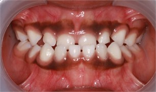
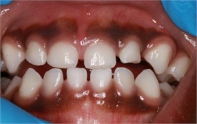
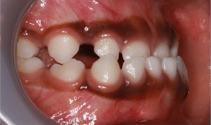
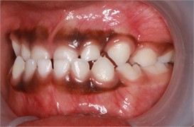
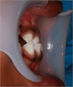
Fig. 1Sibling 1’s initial pictures: a) anterior view in maximum intercuspidation, b) manipulated position of the mandible, c) right-side view, d) left-side view, e) negative overjet
a)
b)
c)
d)
e)
Clinical examination findings: ARM presented with a deciduous dentition with a congenitally missing upper right first deciduous molar (URD). The dental relationship was a mesial step on the molars and Class III on the canines, with an anterior crossbite. The transverse dimensions of the maxilla and mandible were harmonious. A 2 mm lower dental midline deviation was observed, with the chin deviating to the same side. In order to determine the functional component of the anterior crossbite, the patient’s mandible was manipulated into centric relationship, and an edge-to-edge relationship was noted (Fig. 1(b)). The initial dental contact was edge-to-edge on the upper and lower first incisors, which led to a functional forward shift to achieve maximal intercuspidation in centric occlusion. The tongue posture was low and thrusting at rest (Fig. 1).
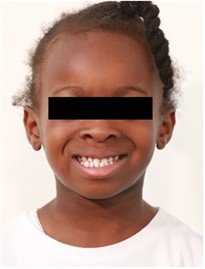
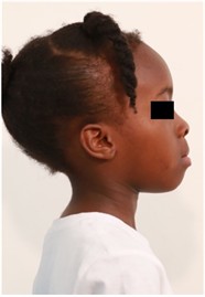
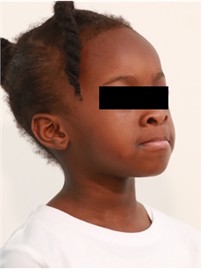
Extra-oral findings: ARM presented in a convex soft tissue profile. The lips were apart mostly with a mouth-breathing tendency. The upper lip was unsupported and thinner than the lower lip. There were no signs or symptoms of a Temporomandibular disorder (Fig. 2).
Radiographic findings: The panoramic and cephalometric X-rays were not obtained due to the parents’ hesitancy about radiation exposure to the child.
Treatment Plan: After the clinical and functional evaluation, the decision was made to apply direct anterior tracks to the upper first incisors to control forward advancement of the mandible. The harmony between upper and lower molar widths and the simplicity of the clinical application were other factors in planning direct anterior tracks.
Fig. 2Sibling 1’s initial facial pictures
a)
b)
c)
Treatment Protocol: The direct anterior tracks on the upper first incisors were planned as the treatment option, using a mock-up technique to reduce chair time.
Laboratory Procedure:
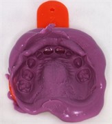
1) Study models were obtained using an alginate impression material, and the manipulated edge-to-edge position of the mandible was registered with a bite registration wax.
2) The working models were mounted on a plane line articulator using the bite registration.
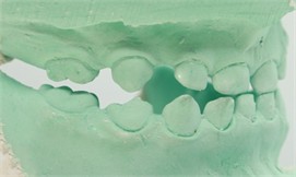
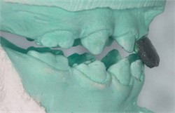
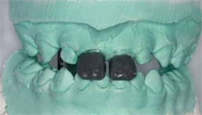
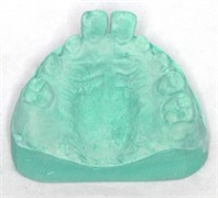
3) The anterior direct tracks were built following the protocol described by Echeverry and Barbosa-Liz [8], using a modelling wax and duplicated.
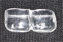
4) Ministar S® compact pressure moulding unit was used to create the mock-up acetate trays following the manufacturer’s instructions (Fig. 3).
Clinical Procedure: Ihe buccal and palatal surfaces of the upper incisors were prepared using a one-step self-etch bonding system (3M ESPE AdperTM PromptTM Self-Etch Adhesive) following the manufacturer’s instructions.
A light-curing hybrid composite (Venus®) was applied to the acetate trays, and each was cured for 40 seconds. The mock-up trays were then removed using gentle pressure and a dental probe.
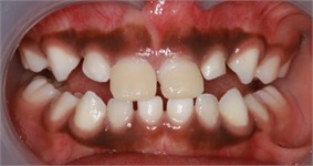
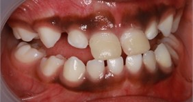
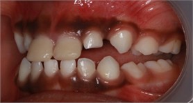
Excess material along the edges was evaluated and finished with 3M™ Sof-Lex™ Discs. Premature occlusal contacts were identified using articulating paper and removed with high-speed finishing bur (Fig. 4.)
Post-op advice: The patient was advised to chew the food on the molars. The parents and the patient were shown the correct tongue posture and were recommended to do a ‘tongue posture’ exercise daily.
Fig. 3Laboratory procedure: a) the working models mounted on the articulator; b)-c) after building the direct tracks with modelling wax; d)-e) duplication and cast model; f) mock-up acetate trays made on duplicated model
a)
b)
c)
d)
e)
f)
Fig. 4Intra-oral picture after the application of anterior direct anterior tracks on the day
a)
b)
c)
Follow-up: Follow-ups are planned at one week, one month, three months, then every three months. During the review sessions, the patient’s complaint, the integrity of the direct track, dental plaque accumulation and the length of the track were evaluated. The inclination of the tracks was assessed and adjusted to maintain the mandibular position behind the maxilla. The tongue posture exercises were reinforced.
Outcomes:
1) The direct tracks remained intact until the exfoliation of the deciduous first incisors.
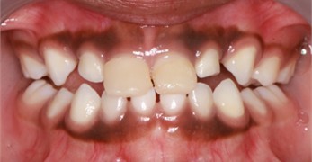
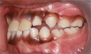
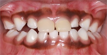
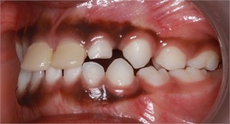
2) A significant improvement in the molar occlusion was observed on the first follow-up session (13 days later) (Fig. 5(a-c)).
3) The patient didn’t attend the first month’s follow-up session.
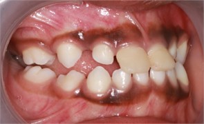
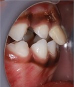
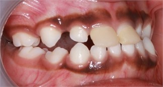
4) A significant improvement in the molar occlusion was observed after three months (Fig. 5(d-g)).
5) The patient reported no signs or symptoms of TMD.
6) The myofunctional exercises were not followed daily. The importance of correct usage of the orofacial muscles for a more stable outcome was reinforced.
Fig. 5a)-c) Follow-up pictures on day 13, d)-g) Follow-up pictures after 3 months
a)
b)
c)
d)
e)
f)
g)
31 Months Follow-up: ARM attended the clinic for a routine check-up after missing follow-ups since the 3rd month (7 years and 2 months old).
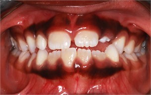
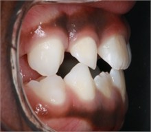
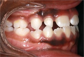
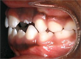
Clinical Examination Findings: The patient presents with early mixed dentition. The molar relationship was Class III on the left and Class I on the right due to mesial drift of the upper right molars, with a 2.5 mm overjet. The tongue posture was low, and the lips were apart mostly (Fig. 6(a-d)).
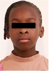
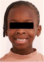
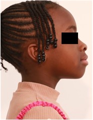
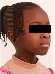
Extraoral Findings: The soft tissue profile was similar compared to the initial images, with more support on the upper lip (Fig. 6(f-i)).
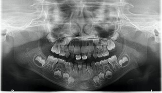
Radiographical Findings: The panoramic -ray taken previously (8 months ago) showed a congenital absence of the upper right first premolar. (Fig. 6(e)).
Fig. 6Follow-up pictures after 31 months: a)-d) intraoral pictures; e) panoramic X-ray, f)-i) facial pictures
a)
b)
c)
d)
e)
f)
g)
h)
i)
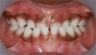
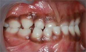
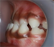
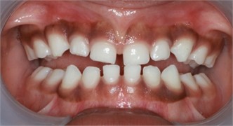
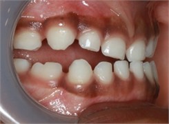
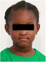
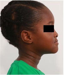
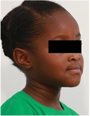
Fig. 7Preop pictures of Sibling 2: a)-c) intraoral pictures in maximum intercuspidation; d)-e) intraoral pictures of the manipulated position of the mandible as described in the text; f)-h) extraoral pictures
a)
b)
c)
d)
e)
f)
g)
h)
2.2. Sibling 2
SM, 4 years 9-month-old girl, the younger sister of Sibling 1, attended the clinic with the same complaint and requested a similar treatment to that of Sibling 1.
Medical history was clear and healthy with no relevant issues. Family history was known as the mother, and the elder sister had the same malocclusion.
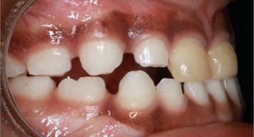
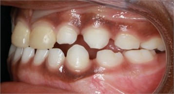
Clinical examination findings: SM presented in deciduous dentition with a mesial step molar relationship and an anterior cross bite. The upper and lower molar widths presented in harmony. The clockwise growth of the premaxilla was noted, associated with an increased overbite of 80 % (Fig. 7(a-c)). The dental midline was coincidental with the facial midline. To determine the functional component of the anterior crossbite, the patient’s mandible was manipulated into centric relationship, and an edge-to-edge relationship was noted (Fig. 7(d, e)). The tongue posture was low and thrusting at rest.
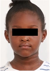
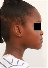
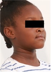
Extra-oral findings: SM presented in a concave soft tissue profile. There were no signs or symptoms of a Temporomandibular disorder (Fig. 7(f-h)).
Treatment Plan: After the clinical and functional evaluation, the decision was made to apply direct anterior tracks to the upper first incisors to control forward advancement of the mandible.
Treatment Protocol: The treatment protocol was followed, as mentioned above. The laboratory and clinical procedures, the post-treatment advice and the follow-up session planning were also the same as in the previous case.
Follow-up: Follow-ups are planned at one week, one month, three months, then every three months. The same follow-up protocol was followed as described in the case of Sibling 1.
Outcomes:
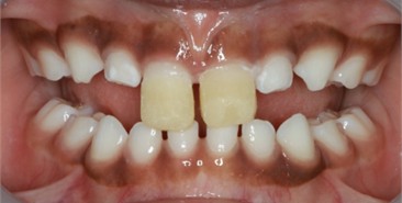
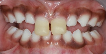
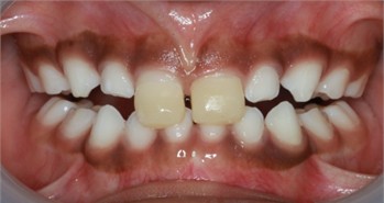
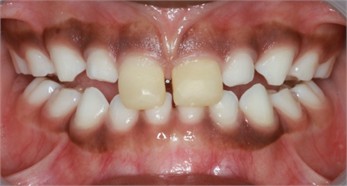
1) A gradual improvement in the molar occlusion was observed during the follow-ups at weeks one, four, and eight (Fig. 8).
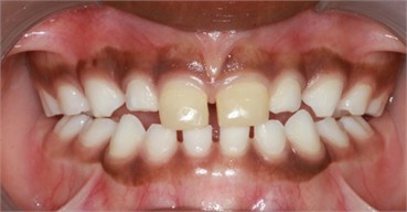
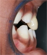
2) The anterior functional crossbite was corrected after one month, and the molar occlusion was near stabilisation in the 7th month. The severity of malocclusion was reduced (Fig. 9).
3) The soft tissue profile improved compared to the initial records, although it remained concave.
4) The tongue posture was low and thrusting towards the lower incisors.
5) The patient reported no signs or symptoms of TMD.
Fig. 8Intraoral pictures of Sibling 2: a) after the application of direct anterior tracks on day 1; b) one week follow-up; c) 4 weeks follow-up; d) 8 weeks follow-up
a)
b)
c)
d)
Fig. 97 months follow-up pictures of Sibling 2: a)-d) Intraoral picture (Note the occlusal contact on the posterior teeth); e)-g) facial pictures
a)
b)
c)
d)
e)
f)
g)
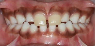
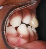
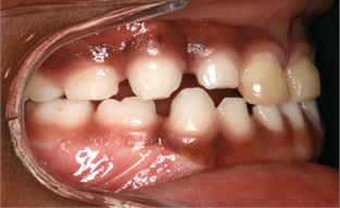
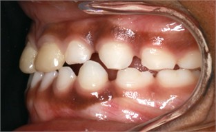
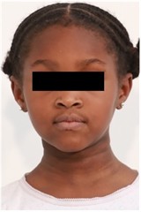
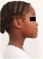
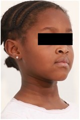
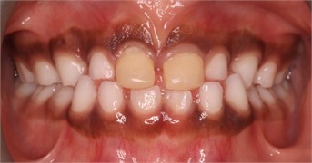
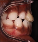
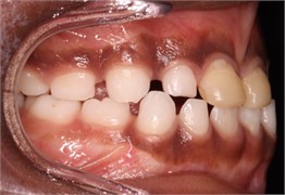
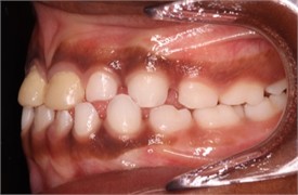
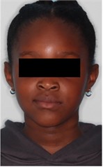
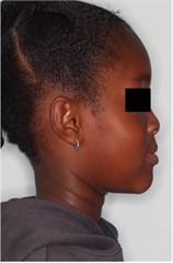
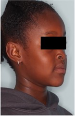
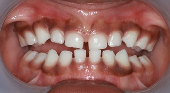
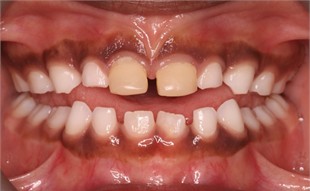
12- and 23-Month Follow-ups: SM attended the clinic for a routine check-up after missing follow-ups since the 2nd month. The direct anterior tracks were intact, and the dental relationship and soft tissue profile had improved (Fig. 10). The lower permanent first incisors erupted with a positive overjet and overbite relationship, and posterior interdigitation had improved, with an acceptable soft tissue profile (Fig. 11). A significant change in the level of the upper anterior gumline was noted due to occlusal levelling (Fig. 12).
3. Discussion
The main objectives of early intervention are to provide a more favourable condition for proper growth and to improve occlusion, function, and facial aesthetics [3, 6, 14]. Anterior crossbite correction is recommended when noted in primary dentition / early mixed dentition using feasible techniques [3, 6]. The most important advantage of early Class III treatment is the reduction in the severity of the discrepancy, which may lessen the complexity of the malocclusion and prevent a dental problem from worsening into a skeletal problem [3, 4, 6].
The preference for the therapeutic tool depends on many factors, such as the patient's age, the severity of the discrepancy, patient compliance, space issues and anchorage needs. The treatment goal is common, which is eliminating the functional forward shift of the mandible and providing a more favourable environment for growth to reduce the need for surgical treatment. This helps eliminate unfavourable stimuli for mandibular growth and the impairment of the maxilla by establishing the correct incisal relationship [3-8].
Fig. 1012 months follow-up pictures of Sibling 2: a)-d) intraoral picture; e)-g) facial pictures
a)
b)
c)
d)
e)
f)
g)
Clinical management studies of Class III cases mostly focus on the mixed/early mixed dentition groups. Early treatment at this age would involve functional appliances (Frankel III or tongue appliance) or orthopaedic devices such as facemasks or chin cups. There isn’t any consensus on the preferred methods because of the complex aetiology of Class III malocclusion and several factors affecting the outcome during treatment [4-6, 12].
The Planas’ Direct Tracks method, following Dr Pedro Planas’ Neuro-occlusal Rehabilitation concept, has been shown to be effective in addressing anterior and posterior crossbite issues in the primary and early mixed dentitions [7, 8, 10, 15-19]. Besides the advantages of the technique's simplicity and the reduced need for patients’ active compliance compared to removable appliances, the Planas’ Direct Tracks redefine the postural morphology of the stomatognathic system due to their 24-hour effectiveness [20].
While the original Planas Direct Tracks were described for posterior teeth, several authors have reported the use of direct anterior tracks on primary incisors for the management of functional anterior crossbite, which creates a barrier to the mandible’s functional anterior shift, leading to a temporary open bite posteriorly and allowing a new posture and occlusal contact between the upper and lower molars [7, 8, 10, 11]. Direct anterior tracks are a modification of the neuro-occlusal rehabilitation approach based on Wilma Simoes’ “determined area” concept, where the upper and lower incisors are in contact on the palato-incisor third of the upper incisor. According to Simoes, this movement opens the “neural incisive circuit”, which may be an indirect way of stimulating the condylar cartilage growth. The maxilla, a secondary aspect, may be influenced sagittally and transversely by the movements of the mandible, and the incisors’ contact may stimulate forward growth [8, 21]. In our case report, both siblings presented with an edge-to-edge bite during the functional examination of the mandible in the centric relationship, supporting the indication for this approach. The direct track built-up was placed on the upper incisors to achieve contact on the palatal surface of the upper incisors.
Fig. 1123 months follow-up pictures of Sibling 2: a)-d) intraoral picture; e)-g) facial pictures
a)
b)
c)
d)
e)
f)
g)
Fig. 12Comparison of the level of the upper anterior gumline between initial and 23-month follow-up pictures: a) initial intraoral picture; b) 23 months later
a)
b)
According to the reports reviewed by Almeida-Junior and Rios, occlusal stability is reestablished between 15 and 45 days after PDT application [10, 14]. In our cases, we have achieved occlusal stability after three months and seven months, respectively. Our findings are similar to Echeverry and Barbosa-Liz’s (six months), who reported that the patient was hyperdivergent Class III with increased mandibular body length [8]. The longer period required to achieve occlusal stabilisation may be associated with the familial Class III tendency observed in both cases; however, this relationship cannot be established from the present report.
The extended follow-up period enabled us to evaluate the impact of early intervention during the early mixed dentition stage, thereby making a significant contribution to the existing literature. We monitored Sibling 1 for 31 months and Sibling 2 for 23 months without any additional interception until the permanent incisors erupted into a positive overjet relationship. In both cases, the intervention eliminated the functional mandibular displacement, established a positive overjet relationship, and reduced the severity of the developing malocclusion. Sibling 1 subsequently received a Simoes Network 3 appliance as part of her ongoing Functional Jaw Orthopaedic treatment, while further Functional Jaw Orthopaedic follow-up was recommended for Sibling 2. As the objective of the present report was to evaluate the outcomes associated with Direct Anterior Tracks, records obtained after the initiation of additional orthopaedic treatment were not included in the analysis. Improvements in occlusal relationships and anterior dentofacial appearance were evident in the follow-up images. These clinical changes are consistent with the principles of Neuro-occlusal Rehabilitation, which proposes that appropriate functional stimuli may contribute to the development of a more balanced stomatognathic system [22]. Both cases in Class III molar relationship demonstrated favourable progress, even after achieving a positive overjet on the incisors and following an extended follow-up period. While the true Class III relationship may not be fully corrected, early intervention is highly advantageous, as Class III tends to stabilise or improve during growth, and treatment outcomes are typically better when addressed promptly [3, 6, 12, 23]. Our findings were similar to the literature, as both cases’ complexity decreased, and they became more manageable by the end of the follow-up period.
4. Conclusion
Within the limitations of this case report, applying Direct Anterior Tracks during deciduous dentition proved effective in early treatment of functional anterior crossbite in two siblings with a familial Class III tendency. The intervention corrected the functional mandibular displacement, established positive overjet and overbite relationships, and these results persisted over follow-up periods of 23 and 31 months. The eruption of permanent incisors into a positive overjet indicates a reduced severity and complexity of the developing malocclusion. Further prospective studies with larger samples are necessary to assess the long-term effectiveness and stability of this method.
References
-
E. H. Angle, “Classification of malocclusion,” Dental Cosmos, Vol. 41, pp. 248–264, 1899.
-
T. D. Foster and M. C. Hamilton, “Occlusion in the primary dentition. Study of children at 2 and one-half to 3 years of age,” British Dental Journal, Vol. 126, No. 2, pp. 76–79, Jan. 1969.
-
“Management of the developing dentition and occlusion in pediatric dentistry. The reference manual of pediatric dentistry,” American Academy of Pediatric Dentistry, 2024.
-
M. A. Hamidaddin, “Optimal treatment timing in orthodontics: A scoping review,” European Journal of Dentistry, Vol. 18, No. 1, pp. 086–96, Feb. 2023, https://doi.org/10.1055/s-0043-1768974
-
S. C. Woon and B. Thiruvenkatachari, “Early orthodontic treatment for Class III malocclusion: A systematic review and meta-analysis,” American Journal of Orthodontics and Dentofacial Orthopedics, Vol. 151, No. 1, pp. 28–52, Dec. 2016, https://doi.org/10.1016/j.ajodo.2016.07.017
-
C. Zhou et al., “Expert consensus on pediatric orthodontic therapies of malocclusions in children,” International Journal of Oral Science, Vol. 16, No. 1, p. 32, 2024, https://doi.org/10.1038/s41368-024-00299-8
-
G. Ramirez-Yanez, “Treatment of anterior crossbite in the primary dentition with esthetic crowns: Report of 3 cases,” Pediatric Dentistry, Vol. 33, No. 4, pp. 339–342, Jul. 2011.
-
J. C. Echeverry and D. Barbosa-Liz, “Direct anterior tracks: Early and functional management of Class III malocclusions-case report and literature review,” Case Reports in Dentistry, Vol. 2019, No. 1, 2019, https://doi.org/10.1155/2019/9323969
-
P. Planas, Rehabilitation neuroclusal. Rio de Janeiro, Brazil: Medsi, 1997.
-
L. A. Almeida-Junior, M. M. Polimeno, A. P. Tortelli, and V. Medau, “Neuroclusal rehabilitation with planas direct tracks in the treatment of anterior crossbite – literature review,” Jaw Functional Orthopedics and Craniofacial Growth, Vol. 4, No. 1, pp. 39–43, Jun. 2024, https://doi.org/10.21595/jfocg.2024.23837
-
L. B. Rossi, K. E. D. C. Pizzol, E. M. Boeck, N. Lunardi, and A. J. I. Garbin, “Correção de mordida cruzada anterior funcional com a terapia de pistas diretas planas: relato de caso,” Revista Da Faculdade De Odontologia De Lins, Vol. 22, No. 2, pp. 45–50, Dec. 2012, https://doi.org/10.15600/2238-1236/fol.v22n2p45-50
-
E. Zere, P. K. Chaudhari, J. Saran, K. Dhingra, and N. Tiwari, “Developing class III malocclusions: challenges and solutions,” Clinical, Cosmetic and Investigational Dentistry, Vol. 10, No. 22, pp. 99–116, 2018, https://doi.org/10.2147/ccide.s134303
-
Y.-R. Zou, Z.-Q. Gan, and L.-X. Zhao, “Clear aligner treatment for a four-year-old patient with anterior cross-bite and facial asymmetry: A case report,” World Journal of Clinical Cases, Vol. 10, No. 15, pp. 5088–5096, 2022, https://doi.org/10.12998/wjcc.v10.i15.5088
-
M. R. Xavier Sancho Rios, A. Alves de Carvalho, A. Silveira Martins, A. Souza Nery, and M. A. Drummond, “Jaw functional orthopedics is a therapeutic alternative for class III malocclusion: a case report,” Jaw Functional Orthopedics and Craniofacial Growth, Vol. 1, No. 2, pp. 44–53, Dec. 2021, https://doi.org/10.21595/jfocg.2021.22059
-
D. A. Lentini-Oliveira, “Planas direct tracks to treat functional crossbites in children: scientific evidence,” Jaw Functional Orthopedics and Craniofacial Growth, Vol. 2, No. 2, pp. 24–31, Nov. 2022, https://doi.org/10.21595/jfocg.2022.22739
-
F. Monte Callado and M. L. Sperandeo, “Anterior crossbite correction in early mixed dentition stage using functional jaw orthopedics principles: a case report with two-years follow-up,” Jaw Functional Orthopedics and Craniofacial Growth, Vol. 1, No. 2, pp. 62–74, Dec. 2021, https://doi.org/10.21595/jfocg.2021.22173
-
A. Devasya, N. K. Ramagoni, M. Taranath, K. E. Prasad, and M. Sarpangala, “Acrylic planas direct tracks for anterior crossbite correction in primary dentition,” International Journal of Clinical Pediatric Dentistry, Vol. 10, No. 4, pp. 399–403, 2017, https://doi.org/10.5005/jp-journals-10005-1473
-
K. Vora, A. Misal, and N. Toshniwal, “An innovative approach for correction of pseudo class III malocclusion with the use of "Planas direct tracks",” APOS Trends in Orthodontics, Vol. 3, No. 6, p. 190, 2013, https://doi.org/10.4103/2321-1407.121440
-
G. Ramirez-Yañez, “Planas direct tracks for early crossbite correction,” Journal of Clinical Orthodontics, Vol. 37, No. 6, pp. 294–298, 2003.
-
A. J. I. Garbin, B. Wakayama, T. A. S. Rovida, and C. A. S. Garbin, “Neuroclusal rehabilitation and planas direct tracks in the posterior crossbite treatment,” RGO – Revista Gaúcha De Odontologia, Vol. 65, No. 2, pp. 109–114, 2017, https://doi.org/10.1590/1981-863720170002000023046
-
W. A. Simoes, “Occlusal plane: A clinical evaluation,” The Journal of Clinical Pediatric Dentistry, Vol. 19, No. 2, 1995.
-
M. Moura, M. Marques, A. C. V. Mello-Moura, and S. F. Ramos, “Neuro-occlusal rehabilitation (NOR) in the early treatment of malocclusion in primary dentition: A scoping review,” Journal of Oral Medicine and Dental Research, Vol. 5, No. 4, pp. 1–22, 2024, https://doi.org/10.52793/jomdr.2024.5(4)-75
-
U. E. M. Schneider-Moser and L. Moser, “Very early orthodontic treatment: when, why and how,” Dental Press Journal of Orthodontics, Vol. 27, No. 2, p. e22spe2, 2022, https://doi.org/10.1590/2177-6709.27.2.e22spe2
About this article

The authors have not disclosed any funding.
The datasets generated during and/or analyzed during the current study are available from the corresponding author on reasonable request.
The authors declare that they have no conflict of interest.
Informed consent for publication was obtained from the legal guardians of both patients who are involved in the case report.

