Abstract
Any dysfunction of vestibular system leads to various diseases, the most common among them is Benign Paroxysmal Positional Vertigo (BPPV). BPPV is most found in elderly people causing vertigo/dizziness which leads to fall and serious injuries. The primary system in inner ear helping in human body balancing and postural adjustment is the cupula. Cupulolithiasis is a type of BPPV caused when otoconia get settles on the cupula. In the present work, a three-dimensional model of cupula and otoconia particle is modelled. A free vibration modal analysis is carried out on the cupula with and without otoconia particle to extract the natural frequency and mode shapes. Change in natural frequency of the cupula due to presence of otoconia particle is presented. Present work has a future scope involving fluid solid interaction (FSI) and extracting response of the cupula incorporating damping to obtain complete dynamic characteristics of cupula. The extracted results help in future investigation of new therapeutical solution to treat BPPV.
1. Introduction
Body balancing is one of the prominent aspects in human daily life. Vestibular system is responsible for balancing of human body. It comprises of otoliths and semicircular canal. Cupula is fixed on crista which is situated in ampulla of semicircular canal senses angular movements. Hair cells present in cupula generates electrical signals which are processed by brain for postural adjustments to maintain body balance. Calcite particles called otoconia gets settled on cupula, causing a disturbance in cupula equilibrium. This condition is referred as a disorder called Benign Paroxysmal Positional Vertigo (BPPV). Studies show the conventional treatment for BPPV (head maneuver) has a long curing time. Predicting dynamic characteristics of cupula helps in better understanding of its behaviour. It leads to future research in introducing new aid to cure BPPV effectively.
Brevern et al., [1] reported the most common vestibular disorder as Benign Paroxysmal Positional vertigo (BPPV) covering approximately 20 %-30 % people in general. Discussed the most common treatment for BPPV as head maneuver, the curing time through this method is usually 2-12 weeks. Squires et al., [2] discussed BPPV as a mechanical disorder of vestibular system and elaborated the discussion about two mechanisms for BPPV namely Cupulolithiasis and Canalithiasis. Several important features emerged from the work is reported. Concluded that the larger cupula displacement as a result of larger or multiple otoconia. Curthoys et al., [3] performed autopsy on infants with fully developed labyrinth to extract horizontal semicircular canal. Analyzed the canal under a microscope and established a cross-sectional model of the canal. Dohlman [4] experimented and described cupula motion during artificially applied endolymph displacement as a motion similar to a wiper-arc. Reported cupula acts as water tight seal between its apex and ampullary wall. Selva et al., [5] reported the value of Young’s modulus for cupula by using bending membrane theory and finite-element methods. They reported the thickness and Poisson ratio of cupula based on several morphological studies. Walther et al., [6] performed energy dispersive X-ray microanalysis (EDX) analysis revealing chemical composition of otoconia and conducted morphological studies to determine size, shape and nature of otoconia.
In the present paper a 3D model of a cupula with and without otoconia is developed. Developed model has a reference of cross section of the semicircular canal. Modal analysis is carried out using Ansys software with appropriate boundary conditions. Natural frequency and mode shapes up to 6th mode are extracted. The change in natural frequency of cupula due to presence of otoconia is analyzed and reported.
2. Modelling of cupula
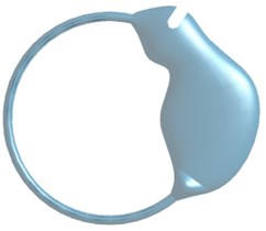
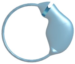
Cupula has circumferential contact along the wall of canal with fixed base end. A 3D Model of the semicircular canal is developed using SolidWorks with appropriate dimensions [3] and is shown in Fig. 1.
Fig. 1Three-dimensional model: a) semicircular canal; b) semicircular canal with cupula
a)
b)
3D model of cupula with thickness 403 µm is obtained by slicing semicircular canal. Front view, side view, top view and isometric view of the obtained cupula is shown in Fig. 2.
The developed cupula has a mass of 3.7557×10⁻⁷ kg.
Fig. 2Principal and isometric views of a cupula

a) Front view
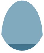
b) Side view

c) Top view
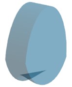
d) Isometric view
3. Modal analysis of cupula
Developed 3D cupula model is imported to Ansys18.1. Modal Analysis is carried out to extract natural frequencies and mode shapes of cupula. A fine tetrahedral mesh is applied for the entire model consisting of 12,805 elements with 21,749 nodes. Fig. 3 shows a cupula with generated mesh.
Cupula is considered as an isotropic elastic solid. Material properties like density, Young’s modulus and Poisson’s ratio considered for the cupula is tabulated in Table 1.
The area of cupula in contact with crista is determined from the model and is considered as fixed boundary condition. Modal analysis is carried out, natural frequencies and mode shapes of cupula are generated and reported in Fig. 4.
Fig. 3Meshed cupula
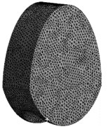
Table 1Material properties of cupula [5]
Parameter | Value |
Density | 1,000 kg/m3 |
Young’s modulus | 5.4×10⁻⁶ N/mm2 |
Poisson ratio | 0.49 |
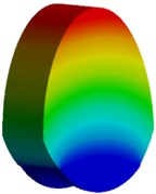
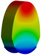
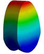
Fig. 4Natural frequency and mode shapes of cupula

a) Mode 1, 4.129 Hz
b) Mode 2, 6.702 Hz
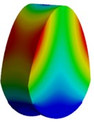
c) Mode 3, 8.935 Hz
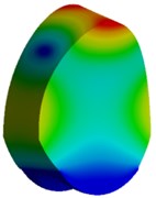
d) Mode 4, 15.343 Hz
e) Mode 5, 17.791 Hz
f) Mode 6, 21.243 Hz
The natural frequency of the cupula is found to be 4.129 Hz, along the flow direction of endolymph at first mode.
4. Modelling of otoconia
Cupula with otoconia on it is modelled. Otoconia are calcium carbonate particles responsible for sensing linear acceleration of head movements in otolith organs [2]. Any harsh or vigorous head movement causes otoconia to sweep into semicircular canal eventually settles on cupula. Presence of otoconia alters the environment and sensing ability of cupula. False signals are sent to brain which is processed to generate false reflexes leading to body imbalance.
Otoconia is a cylindrical bulbous structure with 3+3 rhombohedral planes at 60° at both the ends. The mean size of otoconia is considered as 10 µm [6]. Appropriate solid model of otoconia is developed using SolidWorks 2018 and shown in Fig. 5.
Fig. 53D model of otoconia

5. Modal analysis of cupula with otoconia
Presence of otoconia has an influence on bio-mechanical properties of cupula. This leads to change in stiffness of the system and leading to change in natural frequency.
The material properties of otoconia considered for the analysis is tabulated in Table 3.
Table 3Material properties of otoconia [6]
Parameter | Value |
Density | 2,400 kg/m3 |
Young’s modulus | 6.6 MPa |
Poisson ratio | 0.45 |
Fig. 6a) Cupula with 11 otoconia; b) orientation of 11 otoconia
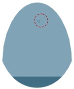
a)

b)
Cupula with otoconia is imported to Ansys18.1. A fine tetrahedral mesh is applied over the entire system of cupula and otoconia consisting of 78,381 elements and 1,18,483 nodes. Fig. 7 shows cupula along with 11 otoconia applied with tetrahedral mesh. Otoconia is considered to have bonded contact with cupula.
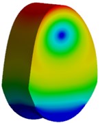
Modal analysis is carried out, natural frequencies and mode shapes of cupula with otoconia are generated. First natural frequency and mode shape is shown in Fig. 8. The change in natural frequency of cupula is insignificant of presence of otoconia.
Fig. 7Cupula – 11 otoconia with mesh
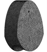
Fig. 8Mode shape of cupula with 11 otoconia, 4.13 Hz
6. Conclusions
In present work, a 3D model of cupula with and without otoconia is developed. Cupula model is generated using the semicircular canal. Appropriate boundary conditions are applied. Modal analysis of both the model is carried out. Natural frequencies, mode shapes are obtained and reported. It is noted that there is no significant change in natural frequency of cupula due to presence of otoconia i.e. natural frequency of cupula without otoconia is 4.129 Hz and natural frequency of cupula with 11 otoconia is 4.13 Hz.
References
-
Von Brevern M., Radtke A., Lezius F., Feldmann M., Ziese T., Lempert T., Neuhauser H. Epidemiology of benign paroxysmal positional vertigo: a population based study. Journal of Neurology, Neurosurgery and Psychiatry, Vol. 78, Issue 7, 2007, p. 710-715.
-
Squires Todd M., Weidman Michael S., Hain Timothy C., Stone Howard A. A mathematical model for top-shelf vertigo: the role of sedimenting otoconia in BPPV. Journal of Biomechanics, Vol. 37, 2004, p. 1137-1146.
-
Curthoys Ian S., Oman Charles M. Dimensions of the horizontal semicircular duct, ampulla and utricle in the human. Acta Otolaryngologica, Vol. 103, 1987, p. 254-261.
-
Dohlman Gosta Some practical and theoretical points in labryrinthology. Proceedings of the Royal Society of Medicine, Vol. 28, 1935, p. 1371-1380.
-
Selva Pierre, Oman Charles M., Stone Howard A. Mechanical properties and motion of the cupula of the human semi-circular canal. Journal of Vestibular Research, Vol. 19, 2009, p. 95-110.
-
Walther Leif Erik, Wenzel Angela, Buder Jana, Bloching Marc Boris, Rudigerkniep, Alexander Blodow Detection of human utricular otoconia degeneration in vital specimen and implications for benign paroxysmal positional vertigo. European Archives of Oto-Rhino-Laryngology, Vol. 271, Issue 12, 2014, p. 3133-3138.
About this article

