Abstract
The Bimler type A utilizes forces derived from muscles, particularly the tongue, concomitantly, it acts as a systemic, dynamic and functional treatment, Bimler appliances transmit neural excitation throughout the system. The objective of this study was to present a clinical case and demonstrate the efficacy of the functional orthopedic appliance, specifically the Bimler A elastic modeler, in the treatment of a patient diagnosed with distoclusion (prognathism of the maxilla and retrognathism of the mandible) according to Bimler and McNamara cephalometry. The case involved a 9-year and 11-month-old male patient with atypical swallowing, respiratory issues, and allergic conditions such as asthma. Clinical examination and complementary tests revealed a large overjet, a narrow maxilla and mandible, an open bite, and distoclusion (retrognathism). The proposed intervention included the installation of the Bimler Elastic Modeler (BEM), type A. The treatment duration was 12 months, with ongoing monitoring every 2 or 3 months. The comprehensive approach, combining BEM type A, chewing exercises, and occlusal adjustments, resulted in orthopedic changes improved mandibular, tongue, lip, and head posture, as well as enhanced chewing balance. Importantly, the appliance effectively repositioned the mandible into a more balanced, normoccluded position without the need for elastic or constant forces. Beyond the orthodontic changes, the active engagement of facial expression muscles during these activities contributed to an overall improvement in facial harmony, achieving lip seal and notable enhancements in breathing. These positive changes extended to the patient’s daily activities and sports performance. The observed outcomes not only increased the child’s active participation in the treatment but also positively impacted self-esteem, driven by the aesthetic, functional, and psychological improvements experienced.
1. Introduction
In 1939, Hans Peter Bimler, a medical professional, was enlisted for wartime service as a doctor. While treating mandibular injuries in soldiers, he innovatively incorporated elastic elements into existing removable appliances. This led to the development of the Bimler Elastic Modeler (BEM), finalized in 1949 [1-4]. Bimler proposed an appliance classification according to the three types of malocclusions: Type A (protruded incisors), Type B (retruded incisors), and Type C (incisors with an inverted relationship) [3-5]. This study reports a case of distoclusion (retrognathism) according to Bimler Cephalometry [4, 5] treated with the functional orthopedic appliance Bimler Elastic Modeler (BEM), type A.
Type A BEM uses forces derived from muscles and movement of the mandible to stimulate transverse development and the shield/tie assembly to reposition the mandible, stimulating growth in length of the mandible [1-4]. Functioning as a systemic, dynamic, and functional treatment [5, 6], BEMs transmit neural excitation throughout the system. Understanding their mode of action necessitates knowledge of embryology and neurophysiology [6, 7]. Functional Jaw Orthopedics, a specialty focusing on functional and growth issues affecting occlusion, aims to remove interferences that are preventing the correct growth and development of the stomatognathic system through guidance, education, occlusal adjustments, and intra-oral appliances [6, 8, 9].
Early intervention in childhood is crucial for detecting functional changes during development [10]. The Stomatognathic System (SS) functions influence jaw development, with malocclusion potentially reflecting functional imbalances impacting skeletal structures and facial muscles. Functional orthopedic appliance seek to balance the stomatognathic system [3, 12, 13]. Treatment with functional orthopedic appliance has a higher likelihood of success, even in cases with an unfavorable prognosis, when diagnosed and intercepted promptly [14].
The sooner the interferences are corrected, through functional guidelines, occlusal adjustments, whether due to wear or additions, or even with the use of appliances, better responses will be obtained, restoring the functionality of the system and correct growth and development [15, 16]. This study presents a case report classified according to Bimler [6] and McNamara Cephalometry, focusing on distoclusion (retrognathism) with large positive overjet, maxillomandibular atresia, and an open bite, treated with BEM type A.
The case underwent a thorough evaluation, study, and diagnosis by the author, adhering to the principles and laws guiding Functional Jaw Orthopedics [5]. All appliances used were crafted by the same orthotechnician, guided and trained based on neurophysiological principles and contemporary manufacturing techniques [5]. The objective was to cause the mandible to change its posture in a Determined Area (DA), promoting a new neural excitation [5]. The time of use of these appliances was 12 months. The case showed satisfactory changes in the sagittal and transverse direction [6-8], which allowed progress in functional orthopedic treatment.
2. Case report
This study delves into the case of Patient Z.K.V, a 9-year and 11-month-old male, seeking care at a private Functional Jaw Orthopedics dental clinic. The anamnesis revealed an uneventful birth, pacifier use until 2.5 years, and the cessation of breastfeeding and bottle-feeding by age 2. Although the overall developmental trajectory was typical, early-life occurrences of shortness of breath, breathing difficulties, restless sleep, snoring, and fatigue were noted. Challenges with attention, shyness, limited engagement in activities, and minimal social interaction were reported during the patient’s school age.
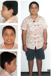
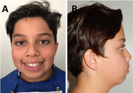
Concerns escalated due to a hereditary predisposition for small jaws, apnea, and snoring within the family. The patient displayed infrequent mastication of hard foods, constant mouth opening, and independent execution of daily activities. The extra-oral clinical examination disclosed atypical swallowing, absence of lip sealing, oral breathing, and a history of allergic conditions such as asthma. A head-forward position was also observed, as illustrated in Fig. 1.
Fig. 1Initial extra-oral photographs of the front, left side, and right side
In the intra-oral clinical examination, no physiological wear was observed. The patient exhibited a narrow maxilla and mandible, Class II division 1 malocclusion (distoclusion) along with large and positive overjet (Fig. 2). In the analysis by McNamara, the patient displayed retrusion of the jaw, with atretic maxilla and mandible, and a relatively small mandible according to the provided table, coupled with an increased Afai (linear distance between the points ENA and Me projected perpendicular to the N-perpendicular). According to Bimler’s analysis, the patient's pattern indicated upper and lower mesoprosopy with a propensity for vertical growth of the maxilla, characterized by retroinclination, a prognathous maxilla, and a retrognathous mandible. Lavergne/Petrovic analysis placed the patient in rotational growth group P1NOB, with a growth potential category at the tissue level 2. Notably, there was a posterior rotation of the mandible, indicating a growth potential smaller than that of the maxilla and an unfavorable prognosis, as depicted in Fig. 3.
Fig. 2a) Initial intra-oral photographs of the patient frontal; b) inferior overjet view; c) lateral overjet view; d) right lateral and e) left lateral
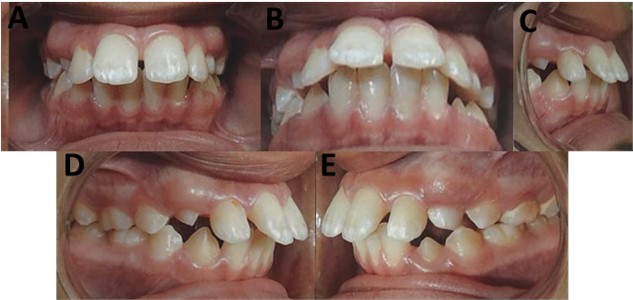
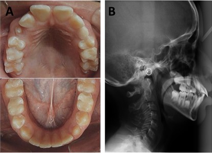
Fig. 3a) Initial occlusal photographs of the maxilla and mandible; b) initial teleradiography
The author conducted a thorough evaluation, study, and diagnosis of this case in accordance with the principles and principles that underpin Functional Jaw Orthopedics [5]. The orthodontic appliance utilized in the study (Fig. 4) was custom-manufactured by an orthotechnician, who received guidance and training rooted in neurophysiological principles and contemporary manufacturing techniques [5]. The primary objective was to induce a change in mandibular posture within a Detemined Area (DA), with the incisal third of the palatal surface of the upper incisors in contact with the buccal incisal third of the lower incisors. This approach aimed to evoke new neural excitation in this altered position [5]. After three months of treatment we can observe changes in occlusion and mandibular advancement (Fig. 4). The duration of appliance use was at 12 months.
Fig. 4Installation Bimler A appliance (A)
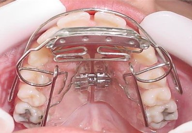
Fig. 5a) Use appliance for 3 months in initial intra-oral photographs of the patient frontal, right lateral and left lateral; b) use appliance for 6 months in initial intra-oral photographs of the patient frontal, right lateral and left lateral
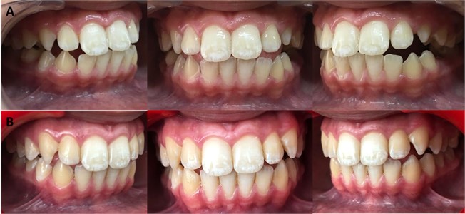
The application of the Bimler A appliance successfully repositioned the mandible into a more balanced alignment, nearing normocclusion. Following the utilization of the Bimler A appliance, a beneficial therapeutic postural adjustment was attained, fostering optimal dynamic functioning of the stomatognathic system. The appliance induced a shift in mandibular posture, accompanied by notable alterations in facial and head posture (Fig. 5).
Fig. 6After 6 months of using the Bimler A appliance
After 6 months of functional orthopedic treatment, the patient exhibited notable improvements in key functions such as breathing, chewing, and swallowing. Additionally, there was a marked enhancement in lip seal (Figs. 6). The treatment also led to favorable adjustments in tongue posture and correction of malocclusion, clearly visible in the accompanying photographs. The orthopedic appliance effectively achieved expansion and alignment, contributing to the observed functional changes. The comprehensive treatment spanned approximately 12 months (Figs. 7). Monthly adjustments were performed on the dorsal arch, frontal laces, Bimler and Coffin arches, with discernible positive changes manifesting over the course of the treatment.
Fig. 7a) Initial intra-oral photographs of the patient frontal and inferior overjet view; b) intra-oral photographs of the patient frontal and inferior overjet view after 12 months of treatment; c) initial occlusal photographs of the maxilla and mandible; d) occlusal photographs of the maxilla and mandible after 12 months of treatment
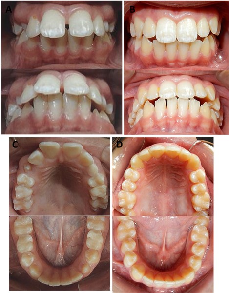
Fig. 8a), b) Changes aesthetic and facials muscles; c), d) radiography changes in growth mandibles
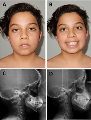
Follow-up appointments take place every 2 months (Figs. 8(a, b)). The reported case has demonstrated commendable changes, facilitating ongoing progress in the treatment. These changes have resulted in a more harmonious masticatory function, fostering optimal dynamic functioning of the stomatognathic system. Notably, the utilization of the orthopedic appliance successfully repositioned the mandible into a balanced and normocclusive alignment, as evidenced in radiographs (Figs. 8(c, d)), without the need for elastic or constant forces [7, 8].
Furthermore, a significant improvement was observed in the patient's self-esteem and social interaction, as reported by their parents (Figs. 7(a, b)).
The case exhibited gratifying changes in both the sagittal and transverse directions [6-8], paving the way for advancements in functional orthopedic treatment.
3. Conclusions
The study compellingly highlighted the appliance’s effectiveness in achieving its intended objective. Functional orthopedic appliance emerge as a promising and efficient alternative for managing distoclusion, particularly when administered during critical periods of growth and development.
References
-
F. J. A. Guila, Ortodontia Teórica e Prática. 2001.
-
T. M. Graber and B. Newmann, Aparatologia Ortodontica Removible. Buenos Aires: Editorial Médica Panamericana, 1979, pp. 337–4801.
-
B. Bimler, “Peter Bimler: uma história de pioneirismo,” Revista Dental Press de Ortodontia e Ortopedia Facial, Vol. 9, No. 6, pp. 18–20, Dec. 2004, https://doi.org/10.1590/s1415-54192004000600004
-
N. Wahl, “Orthodontics in 3 millennia. Chapter 9: Functional appliances to midcentury,” American Journal of Orthodontics and Dentofacial Orthopedics, Vol. 129, No. 6, pp. 829–833, Jun. 2006, https://doi.org/10.1016/j.ajodo.2006.03.019
-
H. P. Bimler and B. Bimler, “Modelador Elástico de Bimler,” Jornal Brasileiro de Ortodontia e Ortopedia Maxilar, Vol. 2, No. 7, pp. 57–64, 1997.
-
W. A. Simões, Ortopedia Funcional dos Maxilares – Através da reabilitação Neuro – oclusal. São Paulo: Artes Médicas, 2003.
-
W. A. Simões, “A study on proprioception and exteroception when Bimler’s, Planas’ and Frankel’s technics are used,” Ortodontia, Vol. 7, No. 2, pp. 153–161, 1974.
-
R. A. Torres, M. H. C. Almeida, T. Wassall, K. J. N. Ferrer, and F. L. C. Bianchini, “Aparelho Ortopédico Funcional de Bimler – Efeitos nas dimensões transversais do arco mandibular,” R.G.O, Vol. 51, No. 4, pp. 317–322, 2003.
-
G. P. Arceguet, “El diagnóstico etiopatogénico en ortopedia funcional de los maxilares,” Rev. Asoc. Argent. Ortop. Funcional Maxilares, Vol. 40, No. 1, pp. 9–20, 2014.
-
C. M. C. T. Maranhão, G. R. P. Santos, C. A. S. Sá, M. F. L. Silva, and S. M. S. Silva, “Paciente pediátrico Classe III de Angle tratado com aparelho funcional regulador de função de Fränkel 3,” Archives of health investigation, Vol. 7, No. 5, 2018.
-
T. Gedrange and W. Harzer, “Muscle Influence on Postnatal Craniofacial Development and Diagnostics,” Journal of Orofacial Orthopedics, Vol. 65, No. 6, pp. 451–466, Nov. 2004, https://doi.org/10.1007/s00056-004-0405-0
-
R. C. B. Bariani, R. Bigliazzi, M. Cappellette Junior, G. Moreira, and R. R. Fujita, “Effectiveness of functional orthodontic appliances in obstructive sleep apnea treatment in children: literature review,” Brazilian Journal of Otorhinolaryngology, Vol. 88, No. 2, pp. 263–278, Mar. 2022, https://doi.org/10.1016/j.bjorl.2021.02.010
-
M. F. F. Silva et al., “Tratamento de mordida aberta anterior em dentadura mista através de aparelho SN6,” Research, Society and Development, Vol. 10, No. 13, pp. 1–11, 2021.
-
G. P. L. Silva, D. G. Andrade, T. R. M. Figueiredo, A. D. Lemos, and M. J. A. L. L. A. Arruda, “Tratamento de Classe III de Angle com tração reversa da maxila e ortopedia funcional dos maxilares: relato de caso,” Archives of Health Investigation, Vol. 7, No. 5, p. 260, 2018.
-
K. E. D. L. Pizzol., C. S. V. Junior, B. A. Almeida, K. B. R. Poquechoque, and V. J. Sanches, “Pistas Indiretas Planas: uma alternativa viável na intervenção precoce de mordida cruzada anterior funcional,” Revista Clínica Ortodontia Dental Press, Vol. 17, No. 1, pp. 78–89, 2018, https://doi.org/10.1002/adma.201703655
-
A. B. Rodrigues, M. M. Uribe, C. A. Morales, and C. H. Martínez-Cajas, “Tratamento precoce de más-oclusões esqueléticas de Classe II – comparação de três aparelhos ortopédicos funcionais: Bionator, Klammt, SN1,” Ortodontia SPO, Vol. 46, No. 6, pp. 558–563, 2013.
About this article

The authors have not disclosed any funding.
The datasets generated during and/or analyzed during the current study are available from the corresponding author on reasonable request.
Márcia do Amaral Sampaio: conceptualization; investigation; methodology, writing – original draft preparation; writing – review and editing. Luciano Aparecido de Almeida-Junior: conceptualiation; investigation; literature review; writing – original draft preparation; writing – review and editing.
The authors declare that they have no conflict of interest.
The Informed Consent Form was presented and signed by those responsible.
