Abstract
In this article, the treatment of two clinical cases is presented, in which the two patients showed distocclusion, severe overbite, required correction of the occlusal plane and movements of the mandible. Both were treated with functional orthopedic appliances In the first case. the patient was 4 years and 5 months old, and in second, 10 years 10 months old. Both had a significant pathological occlusal plane with a large difference in the height between the right and left sides. The lateral movements of the mandible showed completely different Functional Masticatory Angle Planes. The protrusion and opening movements of the mandible also showed deviation. The Functional Orthopedic appliance would advance the position of the mandible forward and would produce better responses if it were able to touch the incisors in the specific area. The forward mandibular position would activate the Ihh (Indian Hedgehog) and produce replication of the mesenchymal cells in the condyle and glenoid fossa. Moreover, it could produce an increase of mandibular length, and regulate the factors of mandibular condylar growth. The space produced between the occlusion as a result of the change to the forward mandible position must stimulate tooth eruption to correct the curve of the occlusal plane. Both cases showed balance of the occlusal plane and same functional masticatory angle Planes. Furthermore, the protrusion and opening movement of the mandible were corrected and no longer showed deviations. Note in both cases that the stability of the mandibular advancement occurred due to maintenance of twice as long to achieve incisor contact.
1. Introduction
Cases of malocclusion (distocclusion) with severe overbite must be treated as soon as it is diagnosed. The early treatment can improve adjustment of development of the maxilla, occlusal plane and balance of the ATM.Furthermore, it produces a physiological balance of the mandibular movements (lateral, protrusion, opening and close) and the muscle system.
Malocclusion (distocclusion) with severe overbite can cause a pathological curve of the occlusal plane. Moreover, a different maxillary curve could develop in the occlusal plane between the right and left sides, when measured from the occlusal plane up to the Camper plane. It is also important to diagnose the changes in the curve of the occlusal plane in the first dentition. The occlusal plane must be flatin primary dentition and slightly curved in the permanent dentition [1]. The occlusal plane is a sagittal curve that will be no more than the sum of the individual occlusal planes of each tooth. These individual planes are called microplanes and formation of the sagital curve arises from their alignment. From canine up to the molar there will be an ascending curve corresponding to the resulting directional muscular forces [2].
The lateral movement of the mandible should have the same masticatory angle according to Planason the right and left sides as well as the same length [3]. The lateral movement should be in lateral-protrusion with the disocclusion in group touching the incisors, canine, pre-molars and molars [3]. The protrusion movement is physiological when the mandible moves forward until it touches the palatal surface, moves downwards, touches the incisal edge and then move forwards and upwards[1]. The protrusion movement is pathological or broken when the mandible moves forward and does not touch the incisors1.Diagnosis of the position of the mandible is very important and the incisor midline should be without deviation in the rest position. Furthermore, when the mandible moves forward in a protrusion movement, it must remain without deviation. There are 3 fundamental principles in Functional Maxillary Orthopedics: neural excitation, change in the posture of the mandible, change in the posture of tongue and change in the therapeutic posture of the mandible [1]. The change in the therapeutic posture of the mandible is when the mandible moves forward until ittouches the incisors in a specific area called Determined Area (DA) [1]. This contact in DA establishes the neuro-muscular system that activates the pterygoid muscle, ATM, condylar cartilage, tongue, temporal and facial muscle. Furthermore, there is a relationship between the following structures: respiratory air space, cervical spine, tongue, head position, mandible, neck, ATM and inframandibular region [4]. This activation of the neural-muscular system and their relationship with the anatomical structures makes it possible to change the position of the teeth and correct the occlusal plane curve. There are three possibilities foradvancing the mandible to correct the distocclusion with overbite and it depends on several factors for example, the position of the mandible, deviation of the incisor midline, type of protrusion movement, development of the ATM, teeth position and others. The first option foradvancing the mandible is by direct pro-translation movement [5] (advance the mandible until it touches the DA), the second option is pro-translation movement in 2 steps [5] (advance the mandible in 2 steps) and the third option is gradual pro-translation movement by interference [5] (pain, overjet greater than 7 mm, caution). The change in posture of the mandible for correcting the distocclusion produces the interocclusal space. It is very important and necessary to balance the curve of the occlusal plane and correctthe position of the teeth.
In research conducted by Petrovic et all [6-8] they found an increase in mitotic activity in the mesenchymal cells of the condyle when the mandible was moved into the forward position. Rabie and Hagg researched the factors regulating mandibular condylar growth at the cellular and molecular levels [9]. Therapy with functional appliance has been found to accelerate and enhance condylar growth [10]. Rabie et all have shown that replication of the mesenchymal cells in the condyle and glenoid fossa couldincreaseby 40-50 % during the forward position of the mandible. The protein Indian Hedgehog (Ihh) is a mechanotrasduction mediator in the condylar cartilage that has shown increased expression with the mandibular in a forward position. This activated the molecular cells to increase the condylar bone formation [11]. Furthermore, the forward position of the mandible is capable of up-regulating the expression of SOX9 and type 2 collagen in the glenoid fossa in addition toincreasing the endochondral ossification of condylar cartilage indicated by expression of type X collagen [12]. The Vascular Endothelial Growth Factor (VEGF) is an angiogenic factor that increasedbone in the posterior glenoid fossa by up to 268%during stepwise mandibular advancement [13]. Furthermore, the stepwise mandibular advancement increasedneovascularization and bone formation in the condyle when a new mandibular advancement began [14]. The transcripcion factor Runx2, increases the protein synthesis in the RNAm in the condylar cartilage during the mandibular advancement [15]. In addition, the mechanical tension increased the protein runx2 in hypertrophic chondrocytes. In adults the mechanical strain leads to condylar growth [16]and the continuous effects of mandibular advancement leads to enhancingmandibular and condylar length [17].
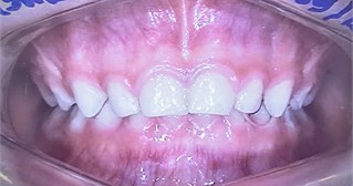
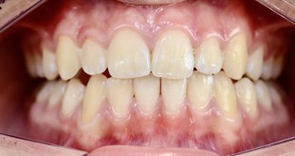
Presentation of the first clinical case concerns the patient, a boy aged 4 years 5 months. In Fig. 1, please note the malocclusion (distocclusion) with overbite, maxillary atresia with the buccal inclination of teeth and deep curve of the occlusal plane.
Bimler's cephalometric analysis showed Superior Profile Angle (N-A) = –1 (Retrognathic), Inferior Profile Angle (A-B) = 19 (Retrogenic), Mandibular Plane (Go-Me) = 32 (Leptognathous), Palatal Plane (Ena-Enp) = –4 (Retro-tilt), Maxillary Incisive Angle = 89 (Retrusion), Interincisive Angle 153 (Bi-retrusion), Mandibular Incisive Angle = 117(Medium), Goniac Angle = 129 (Leptognathous), Dolic/Lepto Dolicognathous.Lavergne/Petrovic Cephalometric Analysis shows SNA = 80 and SNB = 72. Mandible Anterior Rotation (A), Growth Potential Capacity at Tissue Level 2, Mandible smaller than the Maxilla (2) and Mandible Distal Sagittal Relationship (D) with an unfavorable prognosis. Furthermore, curve of the occlusal plane on the left side is higher than that on the right side. The malocclusion was treated with the Bimler Functional Orthopedic appliance, Simões network 1, Simões network 1 with micro-tracks.
Fig. 1Initial front view
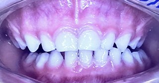
After 3 months, Fig. 2 shows the initial mandibular advancement with contact of the incisors,improved development of the maxilla, and improvedposition of the teeth.
Fig. 2Front view after 3 months
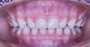
Fig. 3Front view after 6 months
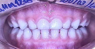
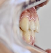
After 6 months, Fig. 3 shows the correction of malocclusion (distocclusion) with overbite and mandibular advancement with incisor contact in DA. Furthermore, improved development of the maxilla and correction of the buccal inclination of teeth were shown. Please, note the remodeling process was taking place and the interocclusal space appeared on the left side, which was larger when compared with the space on the right side. It is also important to reshape the balance of the curve of the occlusal plane curve by tooth eruption and balance of the muscle strength.
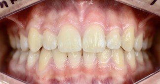
In Fig. 4, at 9 years 11 months, shows maintenance of the advanced mandibular position, incisors in contact and correction of distocclusion with overbite. Furthermore, please note correction of the occlusal plane in the permanent dentition, with a light Spee Curve and the same level of height on both the left and right sides.
Fig. 4Front view after 9 years 10 months
In the Figs. 5, 6 and 7 the profile view showed the initial distal position of the mandible, at 3 months after advancement of the mandible; at6 months after advancement of the position of the mandible with the incisors in contact in DA and correction of distocclusion in the position of the mandible.
Fig. 8 showsmaintenance and stability of the mandibular position at 9 years and 10 months after the patient was treated.
Fig. 5Initial profile
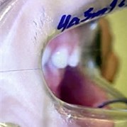
Fig. 6Profile after 3 months
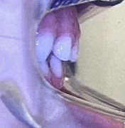
Fig. 7Profile after 6 months
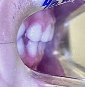
Fig. 8Profile after 9 years 10 months
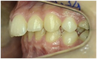
In Fig. 10 note the lateral movement of the mandible showing the latero-retrusion movement on the left side and inFig. 9 the latero-lateral movement on the right side
In Fig. 9 note the lateral movement to the right side showing the latero-lateral movement and in the Fig. 10 note the lateral movement to the left side showing the latero-retrusion movement.
Fig. 9Initial right laterality movement
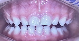
Fig. 10Initial left laterality movement
In Fig. 11 note the lateral movement to right side showing the latero-protrusion movement and in the figure 12 note the lateral movement to the left side showing the latero-protrusion movement. After 6 months treatment, the lateral movement was corrected.
Fig. 11Right laterality movement after 6 months
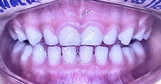
Fig. 12Left laterality movement after 6 months
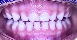
Fig. 13Right laterality movement after 9 years 10 months
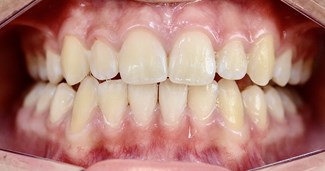
Fig. 14Left laterality movement after 9 years 10 months
In Figs. 13 and 14, with advancement of the mandible, please note the latero-protrusion movement on the right and left sides, respectively, showing improved balance of the curve of the occlusal plane and the same level of the occlusal plane on the right and left sides. Please note the same functional masticatory angle according to Planas, to the right and left sides, thelateral protrusion movement with balance and group disocclusion. Please note the physiological jaw development and the optimal permanent dental position an esthetic and balanced smile.
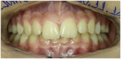
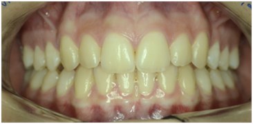
Presentation of the second clinical case concerns the patient, a girl aged 10 years 10 months. Fig. 15, in a front view, note the severe malocclusion distocclusion with severe overbite and left incisor midline deviation; buccal tipping of teeth 12, 21 and 22. Compare the difference between the level of height of the curve of occlusal plane and note that the left side is higher than the right side.
Fig. 15Initial front view of malocclusion
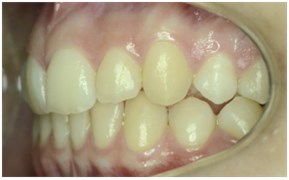
In Figures 16 and 17, right and left sideviews, respectively, please note the severe malocclusion distocclusion with a change in the curve of the occlusal plane. Note the large difference in the position of height between central incisor (higher) and pre-molar (lower).
Fig. 16Initial right side view
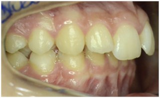
Fig. 17Initial left side view
In Fig. 18, profile side view, note the severe distocclusion with overbite.
Fig. 18Initial profile view
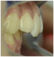
Fig. 19Initial – left laterality movement
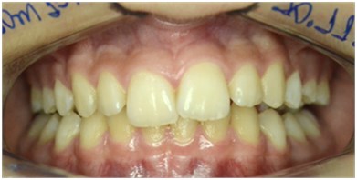
Fig. 20Initial – right laterality movement
In Figs. 19 and 20, respectively, initial of right laterality movement and left laterality movement, note that the Functional masticatory angle according to Planas is smaller on the left side. Furthermore note the lateral-retrusion movement of the mandible on the right and left sides. Teeth position of 43 and 33 are in distal position in relation to teeth position of 13 and 23, respectively. Moreover, the posterior teethdo not touch the molars.
In Fig. 19, please note the small amplitude of movement of right laterality (see the midline incisor position) and compare with the left laterality in Fig. 20. The movement of left laterality has twice the amplitude. This also demonstrates that the movement of mandibular dynamics is performed on the left side.
The movement of protrusion movement showed a deviation of 4 mm to the left side.
Bimler’s cephalometric analysis shows Superior Profile Angle (N-A) = –2 (Retrognathic), Inferior Profile Angle (A-B) = 17 (Retrogenic), Mandibular Plane (Go-Me) = 27 (Mesognathous), Palatal Plane (Ena -Enp) = 0.5 (Orthoinclination), Goniac Angle 122 (Leptognathic), Maxillary Incisive Angle 112 (Medium), Mandibular Incisive Angle 120 (Medium), Meso/Meso Mesognathous.
Lavergne/Petrovic analysis shows SNA = 82.5 and SNB = 76.5. Anterior Rotation of the Mandible (A), Capacity for Growth Potential at Tissue Level 5 and Normal Relation between Jaw and Mandible (1), Distal Sagittal Relation of Mandible (D) with a favorable prognosis.
The malocclusion was treated with Functional Orthopedic appliance: Planas Indirect Tracks, Planas Posterior Track and Planas with Step Posterior Track.
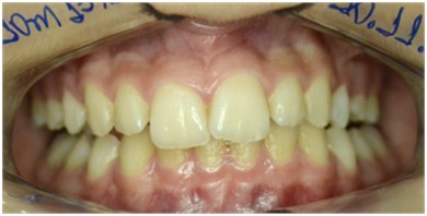
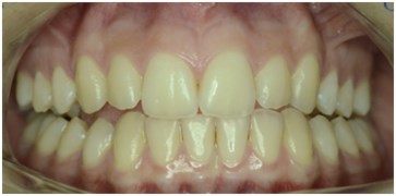
After 3 years 10 months, in the Fig. 21, front view, note the correction of the distocclusion with severe overbite, and also the correction of the mandible position, the incisor midline alignment and the tooth position 12, 21 and 22. Furthermore in Fig. 21, note correction of balance of the curve of the occlusal plane and same the level of height on the right and left sides.
Fig. 21Front view after 3 years 10 months
In Figs. 22 and 23, right and left sideviews, respectively, note correction of the curve of the occlusal plane and correction of difference in the level of height between the central incisor and pre-molar.
Fig. 22Right side view after 3 years 10 months
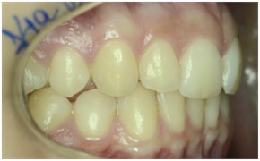
Fig. 23Right side view after 3 years 10 months
In Fig. 24, profile side view, note correction of the distocclusion with overbite and advancement of the mandible with the incisors in contact.
In Figs. 25 and 26, right and left laterality movements, respectively, please note correction of the balance and lateral-protrusion movement with posterior disocclusion in group on the left and right sides. In addition, presents the same Functional Masticatory Angle according to Planas. Furthermore, note the correction of the amplitude in the laterality movement.
Fig. 24Profile view after 3 years 10 months
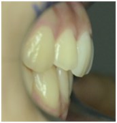
Fig. 25Right laterality movement after 3 years 10 months
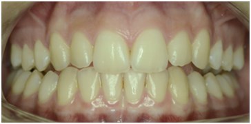
Fig. 26Left laterality movement after 3 years 10 months
The movement of protrusion no longer showed deviation of the midline.
2. Conclusions
Both clinical cases showed balance of the occlusal plane, balance in mandibular movements and excellent esthetic result. When the procedures of correction start at an early age, theyprovide better and faster responses to correcting the malocclusion. Advancement of the mandible can lead to better response to the treatment of malocclusion (distoclusion) with overbite. The Functional Orthopedics appliance stimulated and enhanced the Ihh and factors that regulate condylar growth. The forward advancement of the mandibular position with the incisors in contact in DAled to a better response to the treatment to achieve occlusal plane balance and for correcting the position of the teeth. Furthermore, it stimulated the neural-muscular system to balance the mandibular movements.
References
-
W. A. Simões, Ortopedia Funcional dos Maxilares: Através da Rehabilitacion Neuro-Oclusal. (in Portuguese), Artes Medicas, Divisão Odontológica, 2003.
-
Alonso Albertini Bechelli et al., Occlusion and Diagnosis in Oral Rehabilitation. (in Spanish), 2005.
-
P. Planas, Rehabilitación Neuro-Oclusal. (in Spanish), Medsi, 1994.
-
W. A. Simões, “O octógono da prioridade funcional e a teoria das rédeas musculares,” (in Portuguese), Revista Faculdade de Odontologia UFRGS, 1996, https://doi.org/10.22456/2177-0018.7834
-
W. A. Simões, Artes Médicas. (in Portuguese), 2002.
-
A. G. Petrovic, “Nível de Crescimento tecidual e Potencial de resposta ao tratamento: Rotação de Crescimento e tomada de decisão Terapêutica,” (in Portuguese), Ortodontia, Vol. 22, pp. 32–60, 1989.
-
A. G. Petrovic, J. J. Stutzmann, and J. Lavergne, “Efeito dos aparelhos funcionais sobre a cartilagem do côndilo mandibular,” (in Portuguese), Ortodontia, pp. 64–81, 1991.
-
I. S. Castro, N. A. Campos, A. Assunção, and F. P. A. Lima, “Diferenças interindividuais em teleatendimento de emergências: explicitação por meio da entrevista de autoconfrontação,” (in Portuguese), Revista Brasileira de Saúde Ocupacional, Vol. 31, No. 114, pp. 83–96, Dec. 2006, https://doi.org/10.1590/s0303-76572006000200008
-
A. B. M. Rabie and U. Hägg, “Factors regulating mandibular condylar growth,” American Journal of Orthodontics and Dentofacial Orthopedics, Vol. 122, No. 4, pp. 401–409, Oct. 2002, https://doi.org/10.1067/mod.2002.125713
-
A. B. M. Rabie, T. T. She, and U. Hägg, “Functional Appliance therapy accelerates and enhances condylar growth,” American Journal of Orthodontics and Dentofacial Orthopedics, Vol. 123, No. 1, pp. 40–48, Jan. 2003, https://doi.org/10.1067/mod.2003.45
-
G. H. Tang, A. B. M. Rabie, and U. Hägg, “Indian Hedgehog: A Mechanotransduction Mediator in Condylar Cartilage,” Journal of Dental Research, Vol. 83, No. 5, pp. 434–438, May 2004, https://doi.org/10.1177/154405910408300516
-
A. B. M. Rabie, T. T. She, and V. R. Harley, “Forward Mandibular Positioning Up-regulates SOX9 and Type II Collagen Expression in the Glenoid Fossa,” Journal of Dental Research, Vol. 82, No. 9, pp. 725–730, Sep. 2003, https://doi.org/10.1177/154405910308200913
-
L. Shum, A. B. M. Rabie, and U. Hägg, “Vascular Endothelial growth factor expression and bone formation in posterior glenoid fossa during stepwise mandibular advancement,” American Journal of Orthodontics and Dentofacial Orthopedics, Vol. 125, No. 2, pp. 185–190, Feb. 2004, https://doi.org/10.1016/j.ajodo.2002.12.002
-
Leung and F. Y. C., “Neovascularization and bone formation in the condyle during stepwise mandibular advancement,” The European Journal of Orthodontics, Vol. 26, No. 2, pp. 137–141, Apr. 2004, https://doi.org/10.1093/ejo/26.2.137
-
G. H. Tang and A. B. M. Rabie, “Runx2 Regulates Endochondral Ossification in Condyle during Mandibular Advancement,” Journal of Dental Research, Vol. 84, No. 2, pp. 166–171, Feb. 2005, https://doi.org/10.1177/154405910508400211
-
Xiong and Hui, “Mechanical strain leads to condylar growth in adult rats,” Frontiers in Bioscience, Vol. 10, No. 1-3, pp. 65–73, 2005, https://doi.org/10.2741/1507
-
Xiong H., Hägg U., Tang Gh, Rabie Ab, and Robinson W., “The Effect of Continuous Bite-Jumping in Adult Rats: A Morphological Study,” The Angle orthodontist, Vol. 74, No. 1, pp. 86–92, Feb. 2004, https://doi.org/10.1043/0003-3219(2004)074
About this article

The authors have not disclosed any funding.
The datasets generated during and/or analyzed during the current study are available from the corresponding author on reasonable request.
The authors declare that they have no conflict of interest.
The research met all applicable standards for the ethics of experimentation. Permit to perform biomedical investigation was granted by Dr. Murilo Bovi Corsi, number 46945, 18/10/2022. Participants provided written informed consent prior to the study.
