Abstract
The aim of this study was to assess the dynamic interaction between cardiac cycle duration and pulse wave amplitude during repeated stretching exercises performed before training, after warm-up, and after a 60-minute strength training session. Ten healthy physically active male participants completed a repeated-measures experimental protocol. Cardiovascular signals were recorded in a seated position at three experimental stages: before training, after warm-up, and after training. At each stage, participants first remained seated at rest, after which they performed four bilateral static stretching repetitions, each consisting of 30 s of stretching for each lower limb and separated by 30 s recovery intervals. The interaction between cardiac cycle duration and pulse wave amplitude was evaluated using algebraic data cointegration. The discriminant index was calculated at 10 s intervals, where lower values indicated stronger interaction between the analysed cardiovascular signals. Before training, values demonstrated a phase-dependent decrease-increase pattern, with reductions during stretching repetitions and increases during recovery periods. After warm-up, a similar response was observed during the first two repetitions, but values remained lower during the latter part of the protocol. After the 60-minute strength training session, values decreased at the beginning of the first stretching repetition and remained low throughout most of the remaining protocol. These findings indicate that repeated stretching exercises strengthen the interaction between cardiac cycle duration and pulse wave amplitude, and that this enhanced interaction persists after stretching, particularly under fatigue conditions. This response suggests cumulative effects of exercise and stretching on vago-sympathetic regulation and ventricular–vascular interaction.
1. Introduction
The cardiovascular response to exercise represents a complex dynamic process involving continuous coordination between cardiac and vascular mechanisms to ensure adequate tissue perfusion under changing mechanical, metabolic and autonomic demands. Exercise-induced increases in blood flow are regulated through the interaction of central haemodynamic control and local vascular mechanisms, allowing active tissues to receive sufficient oxygen and substrates during changing workloads [1]. During exercise and recovery, cardiac rhythm, vascular tone and pulse wave characteristics change in an integrated manner rather than as independent physiological responses. Heart rate variability reflects beat-to-beat cardiovascular regulation and is widely used as a non-invasive marker of autonomic modulation [2], [3], while pulse-derived measures obtained from photoplethysmography may provide additional information about peripheral vascular behaviour and pulse-related cardiovascular dynamics [4]. Therefore, the analysis of dynamic relationships between cardiovascular signals may provide more detailed information about cardiovascular regulation than the assessment of separate parameters alone.
Muscle stretching is widely used as part of warm-up, training and recovery routines in sport practice. Previous reviews have suggested that stretching exercise may influence cardiac autonomic regulation, although the direction and magnitude of these effects may depend on the stretching protocol, exercise duration and recovery conditions [5]. During stretching, mechanical deformation of muscle and connective tissue may activate mechanoreceptors and afferent pathways involved in autonomic cardiovascular regulation. Acute passive stretching has been shown to modify heart rate variability responses, with changes in sympathetic and parasympathetic modulation during and after stretching [6]. Similarly, studies investigating post-exercise stretching in athletes have reported changes in HRV-related recovery responses after high-intensity exercise [7]. Since cardiac cycle duration reflects beat-to-beat cardiac control, while pulse wave amplitude is associated with peripheral vascular responses, both parameters can be considered as functionally related components of cardiovascular regulation.
Conventional statistical analysis based only on mean values may not fully describe time-dependent interactions between physiological signals. HRV research has long emphasised that cardiovascular regulation is dynamic and may include both linear and nonlinear properties [3], [8]. More recent work also shows that HRV and pulse rate variability are not identical under all physiological conditions, indicating that pulse-derived signals may contain additional information related to vascular and autonomic responses [9], [10]. For this reason, algebraic data cointegration was used in the present study to assess the dynamic interaction between cardiac cycle duration and pulse wave amplitude during repeated muscle stretching. In this approach, lower values indicate stronger interaction between the analysed signals, whereas higher values indicate weaker interaction and greater divergence in their dynamics. The novelty of the present study lies in the use of algebraic data cointegration to evaluate the time-dependent interaction between cardiac cycle duration and pulse wave amplitude during repeated static stretching performed under different functional states: before training, after warm-up, and after exercise-induced fatigue. While previous studies have mainly examined the effects of stretching on separate heart rate variability or cardiovascular parameters, the present study focuses on the dynamic coupling between cardiac and peripheral vascular signals. This approach provides additional insight into how repeated stretching and prior physical load may modify cardiovascular regulatory interactions over time. The aim of the present study was to assess this interaction before training, after warm-up and after a 60-minute strength training session.
2. Materials and methods
2.1. Participants
The study involved 10 healthy male participants. The mean age of the participants was 20.86±2.03 years, mean height was 184.29±7.13 cm, and mean body mass was 75.00±8.42 kg. Each participant had at least 1 year of regular gym-based training experience and reported at least 3 hours of weekly physical activity, including both aerobic and resistance exercises. All participants were familiarised with the study procedures and testing conditions before the experiment. Exclusion criteria included any diagnosed cardiovascular, neurological, or musculoskeletal disorders; acute injury or pain that could limit exercise performance; use of medication affecting cardiovascular regulation; and participation in strenuous physical activity within 24 hours before testing. The study was conducted in accordance with the Declaration of Helsinki. Ethical approval was granted by the Lithuanian Bioethics Committee, approval No. L-20-1/1, dated 23 January 2020. All participants provided written informed consent before participation.
2.2. Study design
The study employed a repeated-measures experimental design. The same group of 10 healthy male participants completed all testing procedures. Before data collection, participants were familiarised with the study protocol and measurement procedures. Cardiovascular recordings were performed in a seated resting position at three time points: before training, after warm-up, and after training. At each time point, baseline cardiovascular signals were first recorded at rest. Participants then performed repeated muscle stretching exercises, during which cardiovascular signals were continuously monitored.
The warm-up consisted of low- to moderate-intensity running and walking tasks. After the warm-up, participants completed a 60-minute supervised strength training session using resistance-training machines. The session consisted of machine-based strength-endurance exercises targeting the major muscle groups of the lower body, upper body, trunk, and back, including leg press, leg extension, leg flexion, bench press, lat pull-down, abdominal muscle exercises, and back muscle exercises. Exercise intensity was individually prescribed according to standard one-repetition maximum (1RM) assessment and did not exceed 50 % of 1RM. The load was selected to induce muscular activation and exercise-related fatigue rather than maximal strength performance. The session was supervised by a coach and was performed before the final repeated stretching measurement. The primary outcome was the dynamic interaction between cardiac cycle duration and pulse wave amplitude during and after repeated muscle stretching exercises. This interaction was assessed before physical load, after warm-up, and after the 60-minute strength training session.
The dynamic interaction between cardiac cycle duration and pulse wave amplitude was analysed using the algebraic data cointegration method. This approach was used to evaluate cardiovascular functional coherence during stretching under non-fatigued and fatigued conditions.
2.3. Assessment of cardiovascular dynamic interactions
Cardiovascular recordings included cardiac cycle duration and pulse wave amplitude. Cardiac cycle duration was obtained from the R-R interval series recorded using the HRV_Sport system. Pulse wave amplitude was derived from the photoplethysmographic signal recorded by a sensor attached to the left index finger. During each measurement, participants remained seated in a comfortable position, and the same sensor placement and recording conditions were maintained throughout all experimental stages.
At each stage, a 1-min resting baseline was recorded first. Afterwards, participants performed a seated static stretching exercise targeting the posterior thigh and calf muscles of both lower limbs. The exercise was performed in a seated position, with one lower limb extended and the trunk slowly flexed forward toward the foot of the extended limb. Each bilateral stretching repetition consisted of 30 s of stretching for the right lower limb followed by 30 s of stretching for the left lower limb. Four bilateral stretching repetitions were performed, and each repetition was followed by a 30 s passive recovery interval. The stretch was performed actively and maintained to the point of mild discomfort, without pain, bouncing, or forced movements. The same stretching procedure was repeated before training, after warm-up, and after the 60-minute strength training session. Cardiac cycle duration was used as an indicator of beat-to-beat cardiac rhythm regulation, whereas pulse wave amplitude reflected peripheral pulse wave dynamics and vascular response. The obtained cardiac cycle duration and pulse wave amplitude signals were then analysed as paired time series to assess their dynamic interaction during and after repeated stretching exercises.
2.4. Data analysis
Statistical analysis was performed using IBM SPSS Statistics version 26.0 (IBM Corp., Armonk, NY, USA). Data are presented as mean ± standard error of the mean. The normality of data distribution was assessed using the Shapiro–Wilk test. Since the same participants were measured repeatedly, differences between baseline values and each subsequent 10 s interval during stretching and recovery were assessed using the paired Student’s t-test. For each experimental stage, values obtained during repeated stretching and recovery periods were compared with the corresponding baseline values. Because of the exploratory nature of the study and the small sample size, no correction for multiple comparisons was applied. Therefore, the results should be interpreted as indicating time-dependent response patterns rather than isolated effects at individual time points. Statistical significance was set at 0.05.
In addition to conventional statistical analysis, the dynamic interaction between cardiac cycle duration and pulse wave amplitude was assessed using an algebraic data cointegration approach. This method was applied because mean values alone may not fully describe time-dependent cardiovascular interactions during repeated stretching exercises [11].
The recorded cardiovascular signals were analysed as discrete time series and normalised to the [0; 1] interval. For each time point, the difference between paired values of cardiac cycle duration and pulse wave amplitude was calculated. The interaction of neighboring time points was also included in the analysis, and both components were combined into the discriminant index :
where, and represent the normalised values of the analysed cardiovascular parameters at a given measurement point. Lower values indicate stronger dynamic interaction between cardiac cycle duration and pulse wave amplitude, whereas higher values indicate weaker interaction and greater divergence between the dynamics of the two signals. Unlike conventional HRV indices, which mainly quantify variability within cardiac rhythm based on R-R interval dynamics, the index was used to assess the time-dependent coupling between cardiac cycle duration and pulse wave amplitude. Therefore, should be interpreted as a complementary cardiovascular interaction index rather than a replacement for traditional HRV measures. This approach allows evaluation of the dynamic relationship between cardiac rhythm regulation and peripheral pulse wave behaviour during repeated stretching and recovery periods.
In the present study, values were calculated during and after repeated stretching exercises before training, after warm-up, and after the 60-minute strength training session. This approach allowed evaluation of cardiovascular functional coherence under non-fatigued and fatigued conditions.
3. Results
The dynamics of cardiovascular interaction during repeated stretching are presented in Figs. 1-3. values were analysed at 10 s intervals before training, after warm-up, and after the 60-minute strength training session.
Before training, values demonstrated a pronounced phasic pattern throughout the repeated stretching protocol. During the baseline period, values remained relatively stable, with the highest baseline value reaching 0.71±0.10. After the onset of repeated stretching, values decreased during each repetition and increased again during the following recovery periods. The most pronounced decrease was observed during the second repetition, when reached 0.04±0.09, while the lowest values during the first, third and fourth repetitions were 0.09±0.08, 0.05±0.10 and 0.08±0.10, respectively. During recovery periods, values increased again, reaching 0.68±0.09 after the first repetition, 0.57±0.09 after the second repetition, 0.54±0.09 after the third repetition and 0.64±0.10 during the final recovery period. Overall, before training, values changed in a repeated phase-dependent manner, with decreases during stretching repetitions and increases during recovery periods. Significant changes in values were observed during the repeated stretching phases ( 0.05).
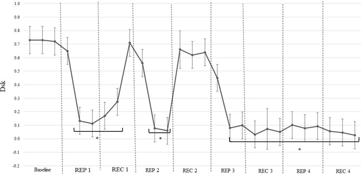
After warm-up, values also demonstrated a phase-dependent pattern during repeated stretching, although the response became less uniform in the later part of the protocol. During baseline, values remained high and relatively stable, reaching 0.73±0.10. After the onset of the first stretching repetition, decreased markedly to 0.12±0.09, followed by an increase during the first recovery period to 0.71±0.10. A similar pattern was observed during the second repetition, where decreased to 0.06±0.10 and subsequently increased during the second recovery period, reaching 0.66±0.14. However, after the second recovery period, values decreased sharply during the third repetition and remained low during the subsequent recovery and fourth repetition phases. In this later part of the protocol, values ranged mainly between 0.03±0.10 and 0.10±0.09, with only small fluctuations between stretching and recovery periods. Significant changes in values were observed during the first and second stretching repetitions, as well as throughout the later part of the protocol from the third repetition to the final recovery period ( 0.05).
Fig. 1Dynamics of Dsk values during repeated stretching performed before training. Note. Dsk, discriminant index; Rep, stretching repetition; Rec, recovery period. * indicates a statistically significant difference compared with baseline values (p< 0.05)
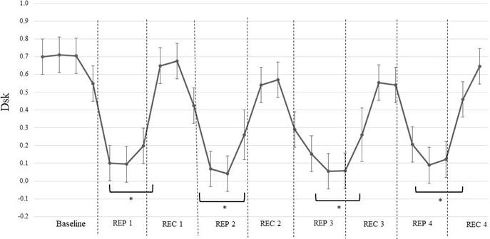
Fig. 2Dynamics of Dsk values during repeated stretching after warm-up. Note. Dsk, discriminant index; Rep, stretching repetition; Rec, recovery period. * indicates a statistically significant difference compared with baseline values (p< 0.05)
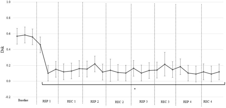
After the 60-minute strength training session, values showed a different time-course pattern compared with the previous conditions. During baseline, values were relatively stable and ranged from 0.56±0.10 to 0.58±0.10. At the onset of the first stretching repetition, decreased markedly, reaching approximately 0.10±0.10. After this initial decrease, values remained low throughout the rest of the protocol, with only small fluctuations between repetition and recovery phases. During the subsequent repetitions and recovery periods, values generally ranged between 0.08±0.12 and 0.21±0.10. The highest value after the initial decrease was observed during the third recovery period, reaching 0.21±0.14, whereas the final recovery period remained low, at 0.11±0.10. Significant changes in values were observed from the first stretching repetition to the final recovery period ( 0.05).
Fig. 3Dynamics of Dsk values during repeated stretching after training. Note. Dsk, discriminant index; Rep, stretching repetition; Rec, recovery period. * indicates a statistically significant difference compared with baseline values (p< 0.05)
4. Discussion
Cardiovascular responses to repeated stretching should be interpreted as part of an integrated cardiac-vascular regulatory process rather than as isolated changes in separate physiological variables. During exercise and recovery, cardiovascular control depends on coordinated adjustments in cardiac rhythm, peripheral vascular tone and blood flow distribution, which together help maintain adequate tissue perfusion under changing physiological demands [1]. In the present study, this coordination was assessed through the dynamic relationship between cardiac cycle duration and pulse wave amplitude using the index. The main finding was that repeated stretching exercises strengthened the interaction between these two cardiovascular signals, as reflected by lower values. Moreover, this enhanced interaction persisted for a certain period after the completion of stretching, particularly after the 60-minute strength training session. The main methodological contribution of this study is that cardiovascular response to stretching was not evaluated only through isolated HRV or pulse-derived parameters, but through the dynamic interaction between cardiac cycle duration and pulse wave amplitude. This allowed us to identify how the strength of cardiac-vascular coupling changed across repeated stretching and recovery periods under non-fatigued and fatigued conditions.
Before training, values demonstrated a clear phase-dependent pattern. They decreased during stretching repetitions and increased again during the following recovery periods. This response suggests that, in the non-fatigued state, repeated stretching induced a transient strengthening of the dynamic relationship between cardiac cycle duration and pulse wave amplitude. However, during recovery intervals, this interaction partially returned toward the baseline level. Such a cyclic decrease-increase pattern may reflect short-term reorganisation of cardiac-vascular regulation during mechanical loading and subsequent recovery. Previous studies have shown that acute stretching can influence heart rate and heart rate variability responses, indicating that even low-intensity stretching may affect autonomic cardiovascular regulation [5], [12]. Therefore, the observed changes before training may be associated with temporary modulation of vago-sympathetic activity and peripheral vascular response during repeated stretching.
After warm-up, the response was partly similar to the pre-training condition during the first two repetitions, as decreases during stretching were followed by increases during recovery. However, after the second recovery period, values remained lower throughout the later part of the protocol. This indicates that warm-up modified the time course of cardiovascular interaction during repeated stretching. Low- to moderate-intensity running and walking may increase cardiovascular readiness by changing autonomic tone, vascular conductance and muscle blood flow. In this condition, repeated stretching appeared to produce a more sustained interaction between cardiac cycle duration and pulse wave amplitude. Stretching-induced autonomic responses have been reported to depend on stretching volume, flexibility level and recovery conditions [6], while post-exercise stretching may also influence HRV-related recovery responses [7]. Thus, the sustained lower values after warm-up may reflect a more persistent coordination between cardiac and vascular components during the later repetitions and recovery phases.
The most pronounced change was observed after the 60-minute strength training session. In this condition, values decreased at the beginning of the first stretching repetition and remained low throughout most of the remaining protocol, with only small fluctuations between stretching and recovery periods. This response differed from the pre-training condition, where recovery intervals were accompanied by a more evident increase in values. The persistence of low values after training suggests that exercise-induced fatigue prolonged the strengthened interaction between cardiac cycle duration and pulse wave amplitude. Since heart rate variability is strongly influenced by autonomic regulation, physical load and recovery processes [3], [8], [10], the sustained cardiac-vascular coupling observed after training may indicate a cumulative effect of strength exercise and repeated stretching on cardiovascular regulation.
The physiological basis of these findings may be related to the combined influence of autonomic and peripheral vascular mechanisms. During stretching, mechanical deformation of muscle and connective tissue can activate mechanosensitive afferent pathways involved in cardiovascular regulation. Passive stretching has been used as a model for mechanoreflex activation and has been shown to evoke cardiovascular and autonomic responses in young adults [13]. Cardiac cycle duration reflects beat-to-beat cardiac regulation, whereas pulse wave amplitude is influenced by peripheral vascular behaviour and pulse wave dynamics. Importantly, pulse-derived measures are not always equivalent to heart rate variability and may be affected by vascular and haemodynamic factors [4], [9]. Therefore, the strengthened relationship between cardiac cycle duration and pulse wave amplitude may reflect not only changes in vago-sympathetic regulation, but also modifications in ventricular–vascular interaction.
The use of algebraic data cointegration provided additional information about cardiovascular regulation by allowing the interaction between two physiological signals to be assessed over time. Conventional HRV analysis provides valuable information about autonomic modulation, but cardiovascular regulation also includes dynamic and nonlinear properties that may not be fully captured by mean-based parameters alone [3], [8]. In the present study, lower values indicated stronger dynamic interaction between cardiac cycle duration and pulse wave amplitude, whereas higher values indicated weaker interaction or greater divergence between the two signals. Therefore, the persistence of lower values after repeated stretching, especially under post-training fatigue conditions, suggests that physical load may prolong the modification of cardiac-vascular interaction.
Overall, the present findings indicate that repeated stretching exercises induce time-dependent changes in the dynamic relationship between cardiac cycle duration and pulse wave amplitude. Before training, this response was mainly transient and phase-dependent, whereas after warm-up and especially after strength training it became more sustained. These results suggest that the cardiovascular response to stretching depends on the functional state of the body and may be influenced by prior physical load and fatigue. This supports the view that stretching performed after exercise is not only a musculoskeletal intervention, but also a stimulus capable of modifying cardiovascular regulatory interactions.
This study has several limitations. First, the sample size was relatively small and included only healthy physically active young men; therefore, the findings cannot be directly generalised to women, older individuals, or clinical populations. Second, the study focused on acute cardiovascular responses to repeated stretching, and longer-term adaptations were not assessed. Finally, the index reflects dynamic interaction between cardiovascular signals, but it does not allow direct identification of the underlying autonomic or vascular mechanisms.
5. Conclusions
Repeated stretching exercises strengthen the interaction between cardiac cycle duration and pulse wave amplitude, with this enhanced relationship persisting for a certain period after the completion of stretching. These cardiovascular responses suggest cumulative effects of exercise and stretching on vago-sympathetic regulation and ventricular-vascular interaction.
References
-
M. J. Joyner and D. P. Casey, “Regulation of increased blood flow (hyperemia) to muscles during exercise: A hierarchy of competing physiological needs,” Physiological Reviews, Vol. 95, No. 2, pp. 549–601, Apr. 2015, https://doi.org/10.1152/physrev.00035.2013
-
Task Force Of the European Society of Cardiology, The North American Society of Pacing, and Electrophysiology, “Heart rate variability: standards of measurement, physiological interpretation, and clinical use,” Circulation, Vol. 93, No. 5, pp. 1043–1065, 1996, https://doi.org/10.1161/01.cir.93.5.1043
-
F. Shaffer and J. P. Ginsberg, “An overview of heart rate variability metrics and norms,” Frontiers in Public Health, Vol. 5, Sep. 2017, https://doi.org/10.3389/fpubh.2017.00258
-
E. Mejía-Mejía, J. M. May, R. Torres, and P. A. Kyriacou, “Pulse rate variability in cardiovascular health: A review on its applications and relationship with heart rate variability,” Physiological Measurement, Vol. 41, No. 7, p. 07TR01, Jul. 2020, https://doi.org/10.1088/1361-6579/ab998c
-
A. Wong and A. Figueroa, “Effects of acute stretching exercise and training on heart rate variability: A review,” Journal of Strength and Conditioning Research, Vol. 35, No. 5, pp. 1459–1466, 2021, https://doi.org/10.1519/jsc.0000000000003084
-
E. D. C. Rubini, P. T. V. Farinatti, C. C. Diano, M. J. Passos Bento Vianna de Oliveira, and E. B. Da Silva, “Effect of single and multiple passive stretching exercises upon heart rate variability in individuals with high and low flexibility levels,” Human Movement, Vol. 22, No. 3, pp. 11–18, Feb. 2021, https://doi.org/10.5114/hm.2021.100320
-
Y. Yıldırım, R. Arabacı, A. K. Güngör, and R. Görgülü, “The effects of dynamic and static stretching exercises performed to elite wrestlers after high intensity exercise on heart rate variability,” (in French), Science and Sports, Vol. 38, No. 1, pp. 2–11, 2023, https://doi.org/10.1016/j.scispo.2022.03.008
-
G. E. Billman, “Heart rate variability? a historical perspective,” Frontiers in Physiology, Vol. 2, 2011, https://doi.org/10.3389/fphys.2011.00086
-
E. Mejía-Mejía, K. Budidha, T. Y. Abay, J. M. May, and P. A. Kyriacou, “Heart rate variability (HRV) and pulse rate variability (PRV) for the assessment of autonomic responses,” Frontiers in Physiology, Vol. 11, Jul. 2020, https://doi.org/10.3389/fphys.2020.00779
-
S. Sammito, B. Thielmann, and I. Böckelmann, “Update: factors influencing heart rate variability-a narrative review,” Frontiers in Physiology, Vol. 15, 2024, https://doi.org/10.3389/fphys.2024.1430458
-
L. Bikulčienė, E. Venskaitytė, L. Gargasas, and V. Jurkonis, “The measure of human vital signals complexity by matrix analysis,” in Chaos and complex systems, pp. 449–458, 2012, https://doi.org/10.1007/978-3-642-33914-1_63
-
P. T. Farinatti, C. Brandão, P. P. Soares, and A. F. Duarte, “Acute effects of stretching exercise on the heart rate variability in subjects with low flexibility levels,” Journal of Strength and Conditioning Research, Vol. 25, No. 6, pp. 1579–1585, Jun. 2011, https://doi.org/10.1519/jsc.0b013e3181e06ce1
-
G. C. S. Lehnen et al., “Cardiovascular and autonomic responses in young adults during and after mechanoreflex activation via passive forearm stretching: are there sex differences,” Journal of Applied Physiology, Vol. 139, No. 5, pp. 1282–1292, 2025, https://doi.org/10.1152/japplphysiol.00645.2025
About this article

The authors have not disclosed any funding.
The authors thank the participants who took part in the study and the staff of Lithuanian Sports University for their assistance during testing.
The datasets generated during and/or analyzed during the current study are available from the corresponding author on reasonable request.
Živilė Kairiūkštienė: conceptualization, methodology, formal analysis, validation, resources, project administration, supervision, writing-original draft preparation, writing-review and editing. Aistė Unskinaitė: investigation, data curation. Dominykas Tvardauskas: investigation, data curation, formal analysis, software, visualization, writing-original draft preparation. Kristina Poderienė: conceptualization, methodology, software, validation, resources, supervision, writing-review and editing.
The authors declare that they have no conflict of interest.
The research adhered to all applicable laws and ethics standards and was conducted in accordance with the Declaration of Helsinki. Permission to perform the research was granted by the Lithuanian Bioethics Committee, No. L-20-1/1, 23 January 2020. Participants provided informed consent prior to the research.
Not applicable. This study was not registered as a clinical trial because it was conducted as an exercise physiology study involving standardised physical exercise and repeated stretching tasks in healthy participants. No drugs, medical devices, invasive procedures, clinical treatments, or clinical outcomes were involved; therefore, clinical trial registration was not required.

