Abstract
Cardiovascular diseases are the leading cause of death among people up to 65 years old. One of the most common cardiovascular disease is thrombosis. To remove the blood clot inside the artery, advanced invasive devices are needed. Nevertheless, medicine of today is not technically fully ready to cope with the challenges. Because of that, tube-shaped ultrasound waveguide wire was designed and the main parameters, such as resonant frequency for displacement towards axis were examined.
1. Introduction
Heart and cardiovascular diseases, related to iliac, femoral and radial artery pathologies, might appear because of the thrombus. Incensement of more and more cases of diseases such as obliterative atherosclerosis, obliterative thromboangiitis, etc. is noticed. At the moment these pathologies are being cured by huge spectra of various ways of treatment, developed to restore the blood flow inside the artery, using invasive fermentative and mechanical thrombus removal tools.
Nevertheless, current treatment methods are limited due to common surgery complications. There is a clear need for the newer, alternative, and less risky and hazardous ways of arterial thrombosis treatment.
During the last few years, ultrasonic methods, which are the most perspective among restoring blood flow in the artery, affected by arterial thrombosis, attracts more and more attention. It is related to the increasing number of researches in the field of ultrasound impact human body tissues. Recent researches [1-3] showed that low frequency and intensity ultrasound increases elasticity of the damaged artery walls. Researches with the same or similar conclusions was performed by other authors as well [4].
2. Current solutions
Extensive review of current invasive arterial thrombosis treatment tools can be seen in the Fig. 1. After the invention of ultrasonic resonator in 2003, few other ultrasound systems were developed. Most noticeable is show in Fig. 1(d). This system has the option to transfer the limited amount of drugs to the spot where arterial thrombosis caused clot appeared. Despite the novelty of the design, it is limited due to volumetric limitation and durability problems of the spring.
The research showed that it is needed to design ultrasound blood vessels cleaning device which is able to:
1) Work in invasive mode, as it is recognised to be the most effective;
2) Be able to provide needed amount of drugs to the cleaning point;
3) Be able to instantly suck the scurf of blood clot at the same time;
4) To provide adequate displacement forward (on axis).
Fig. 1Current patented ultrasound wave guide wire construction, where: a) ultrasonic resonator [5], b) steerable ultrasound catheter [6], c) ultrasound wave guide wire for internal blood vessels cleaning [7], d) ultrasound device for inner blood vessels cleaning [8]
![Current patented ultrasound wave guide wire construction, where: a) ultrasonic resonator [5], b) steerable ultrasound catheter [6], c) ultrasound wave guide wire for internal blood vessels cleaning [7], d) ultrasound device for inner blood vessels cleaning [8]](https://static-01.extrica.com/articles/17562/17562-img1.jpg)
a)
![Current patented ultrasound wave guide wire construction, where: a) ultrasonic resonator [5], b) steerable ultrasound catheter [6], c) ultrasound wave guide wire for internal blood vessels cleaning [7], d) ultrasound device for inner blood vessels cleaning [8]](https://static-01.extrica.com/articles/17562/17562-img2.jpg)
b)
![Current patented ultrasound wave guide wire construction, where: a) ultrasonic resonator [5], b) steerable ultrasound catheter [6], c) ultrasound wave guide wire for internal blood vessels cleaning [7], d) ultrasound device for inner blood vessels cleaning [8]](https://static-01.extrica.com/articles/17562/17562-img3.jpg)
c)
![Current patented ultrasound wave guide wire construction, where: a) ultrasonic resonator [5], b) steerable ultrasound catheter [6], c) ultrasound wave guide wire for internal blood vessels cleaning [7], d) ultrasound device for inner blood vessels cleaning [8]](https://static-01.extrica.com/articles/17562/17562-img4.jpg)
d)
3. Design of the tube-shaped ultrasound wave guide wire
Tube shaped ultrasound wave guide wire with the holes in the working end of it was offered as the alternative to the current patented thrombosis treatment solutions.
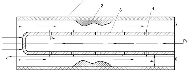
Fig. 2Ultrasonic blood vessels cleaning device (cross-section)
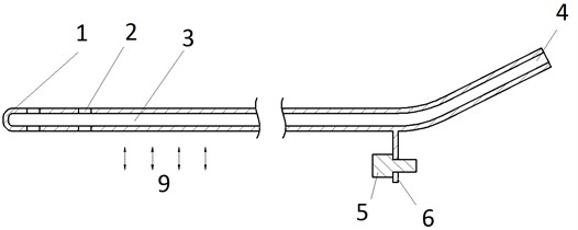
Fig. 3Ultrasonic blood vessels cleaning system, where: 1) wall of the waveguide, 2) waveguide hole, 3) channel, 4) intake, 5) screw, 6) screw hole, 7) concentrator, 8) transducer
The wave guide wire is treated as medical device and can be used to clean the inner walls of the artery, as well as the tool to destroy the thrombus and suck out the scurf of the blood clot at the same time.
To be able to provide a sufficient quantity of drugs to the needed place of the artery, tube-shaped wave guide wire was chosen. The drilled holes at the end of it works both, as intake and suction hole if needed.
Adequate feed of the drugs to the damaged place of the blood vessel is secured by unique design the of wire. The lug which allows the device to get attached to the concentrator is separated from the tube, through which the drugs are delivered. The construction is explained in Fig. 3.
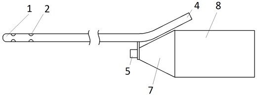
Fig. 4Liquid movement via the wave-guide wire. 1) wall of the blood vessel, 2) ocludator, 3) tube-shaped wave guide wire, 4) hole at the end of wave guide wire
Such construction of the wave guide wire allows to affect the ocludator not only mechanically but also by the flow of physiological fluid, provided through the intake, marked as number 4 in Fig. 3.
4. Experimental research
To determine resonance frequency for the biggest displacement toward z axis, the experiments on the Polytec experimental table were carried on.
Fig. 5Scheme of experiment: 1) polytec machine (laser source), 2) waveguide, 3) holder, 4) anti-vibration table
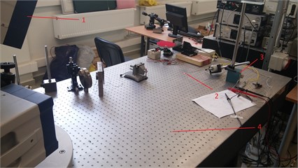
Fig. 6Determining the resonance frequency for the best displacement on z axis (frequency 23.1904 kHz, magnitude: red – 22.843 nm, green – 14.929 nm, blue – 21.035 nm)
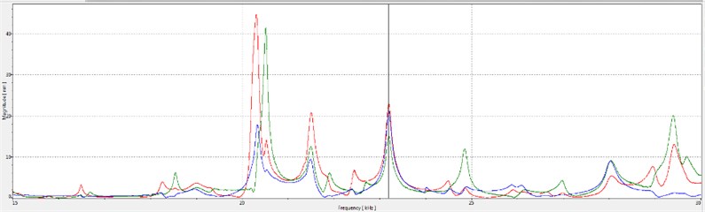
The resonance frequency of 23,19 was obtained, providing biggest 7,02 micrometer displacement towards axis.
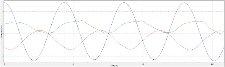
Fig. 7x, y, and z axis displacement on resonant requency (time 43 μs, displacement: red –2.9499 μm, green – 2.2656 μm, blue – 7.0243 μm)
5. Conclusions
Unique design ultrasound invasive blood vessels cleaning device was constructed. It allows not only to suck out the scurf of the just destroyed blood clot but also provide the needed amount of medicine to the needed spot of the damaged blood vessel.
Resonance frequency of 350 mm length tube-shaped wave guide wire is 23,1904 kHz. During the resonance, the biggest displacement towards axis, 7,02 μm is determined.
References
-
Bubulis A., Jurėnas V., Minchenia V. T., Stepanenko D. A., Bobrovskaja A. I., Chizh D. V. Study of the process of interaction between low-frequency ultrasound and biological tissue phantoms. Journal of Vibroengineering, Vol. 13, 2011, p. 586-589.
-
Bubulis A., Jurėnas V., Stepanenko D., Minchenya V. Effect of design of the distal part of wave guide concentrator on efficiency of destroying blood clots during ultrasound angioplasty. “Science and Education” International Scientific Seminar, 2011, p. 11-14.
-
Tun C. Effektivnost Vosstanovlenija Prochodimosti Porazhennix Aterosklerozom Arterij Ultrazvukovimi Volnovodami Razlichnich Modofikacij in Vitro. BelMAPO, 2006, p. 21.
-
Siegal R. J., Gunn J., Ahsan A., Fishbein M. C., Bowes R. J., Oaukley D., Wales C., Steffen W., Campbell S., Nita H. Use of theraupeutic ultrasound in percutaneous coronary angioplasty. Experimental in vitro studies and initial clinical experience. Curculation, 1994, p. 1587-1592.
-
Peterson T., Pal D. Ultrasonic Resonator. US Patent No. US6617760, 2003
-
Nita H., Sarge J. Steerable Ultrasound Catheter. US Patent No. US7335180, 2008
-
Bansevičius R., Bubulis A., Jurėnas V., Minchenia V., Valaika M. Ultrasound Wave Guide Wire for Internal Blood Vessels Cleaning. EU Patent No. EP2065002, 2009
-
Bubulis A., Navickas J., Mažeika D., Janušas G., Naginevičius V., Ostaševičius V., Palevičius A. The Ultrasonic Cleaner of the Internal Blood Vessels – Ultragarsinis vidinio kraujagyslių valymo įrenginys. LT Patent Application No. 2015 048, 2015.
About this article

This research is funded by Research Council of Lithuania, Project No. MIP-097/15.
