Abstract
This paper presents the design and preliminary structural assessment of a cable-driven elbow exoskeleton, AlmatyExoElbow, intended for upper-limb rehabilitation. The proposed system employs a modular architecture to improve biomechanical compatibility while reducing distal segment mass. The exoskeleton provides two degrees of freedom, including flexion-extension and forearm pronation-supination. A cable-driven mechanism enables remote actuator placement and reduces parasitic loading associated with mechanical-anatomical axis misalignment. A 3D CAD model was developed and evaluated using finite element analysis in SolidWorks Simulation. Stress, displacement, and strain distributions were analyzed to identify critical structural regions and guide future design optimization. The results provide an initial assessment of the proposed concept and support further development of rehabilitation-oriented elbow exoskeleton systems.
1. Introduction
Upper-limb exoskeletons have gained increasing attention for rehabilitation after stroke, injuries, and neuromuscular disorders [1-4]. Conventional therapy often relies on repetitive therapist-assisted exercises, which may suffer from inconsistency and limited training intensity [5]. Robotic rehabilitation systems offer a promising approach for improving movement recovery and therapy efficiency [6].
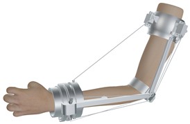
The elbow joint is essential for daily activities and enables flexion-extension and forearm pronation–supination motions (Figs. 1-2) [7-9]. Due to the complex biomechanics of the elbow, misalignment between anatomical and mechanical axes may generate parasitic loads and reduce user comfort [9]. Cable-driven mechanisms have been increasingly adopted in rehabilitation devices because they allow remote force transmission, reduced distal mass, and improved adaptability [10-14].
Although various rehabilitation exoskeletons have been reported, many systems remain mechanically complex and difficult to adapt as lightweight wearable devices [12-18]. Therefore, this work presents AlmatyExoElbow, a lightweight 2-DOF cable-driven elbow exoskeleton intended for rehabilitation applications.
Unlike our previous studies focused on numerical evaluation of rehabilitation devices [19-20], the present work concentrates on the conceptual mechanical design and preliminary structural assessment of the proposed system. Finite element analysis is used to identify critical stress and deformation regions and to provide guidance for future structural optimization. The proposed design aims to improve biomechanical compatibility, reduce distal segment mass, and support rehabilitation-oriented elbow movements.
2. Mechanical design and working principle
2.1. System architecture
The proposed AlmatyExoElbow is a lightweight modular exoskeleton designed for elbow rehabilitation. The design aims to improve biomechanical compatibility, reduce parasitic loads, and enhance adaptability during rehabilitation exercises.
The system consists of a proximal module, a distal forearm module, linkage elements, and a cable-driven transmission mechanism. Remote actuator placement reduces distal segment mass and inertia, while the modular architecture facilitates adjustment for different users and simplifies maintenance.
Fig. 1 shows the exploded view of the proposed design. The main components include: (1) proximal ring module, (2) central joint housing, (3) elbow ring, (4) linkage bars, (5) protective housing, and (6) actuator mounting unit. These elements provide structural support and force transmission between the arm and forearm segments.
The current configuration represents a preliminary conceptual prototype intended for initial biomechanical and structural assessment. Finite element analysis was used to identify critical stress concentration regions that will guide future structural reinforcement and geometric optimization. Compared with conventional rigid rehabilitation devices, the proposed cable-driven architecture offers improved adaptability and reduced mechanical constraints.
Fig. 1Exploded view of the elbow exoskeleton
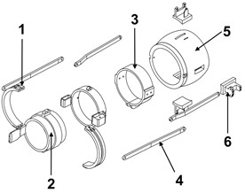
Fig. 2Elbow exoskeleton mounted on the arm
2.2. Structural modules and human-interface design
The exoskeleton employs ring and semi-ring modules to improve load distribution, biomechanical compatibility, and user comfort during rehabilitation exercises. As shown in Fig. 2, the proximal module serves as the main support structure and incorporates the actuator mounting and cable-routing elements, while the distal module is attached to the forearm and transfers assistive rotational forces.
Adjustable fastening elements allow adaptation to different user anatomies and provide stable fixation during movement. The cable-driven architecture enables remote force transmission, reducing distal segment inertia and improving motion flexibility compared with conventional rigid rehabilitation devices.
The modular and detachable design simplifies installation, maintenance, and future modifications. The current prototype represents a preliminary rehabilitation-oriented configuration intended for initial biomechanical and structural assessment, while further optimization and experimental validation are planned for future work.
2.3. Cable-driven mechanism and working principle
The proposed exoskeleton employs a cable-driven mechanism to transmit forces from the actuator unit to the forearm module. Remote actuator placement reduces distal segment mass and inertia, improving biomechanical compatibility during rehabilitation movements.
Fig. 3 illustrates the kinematic scheme of the system. The mechanism uses cable routing through guide elements and attachment points (A, B, C). Variations in cable lengths (–) generate rotational motion of the distal module and assist elbow flexion–extension and forearm pronation–supination movements.
Compared with rigid transmission systems, the cable-driven architecture reduces alignment requirements between anatomical and mechanical joint axes, thereby decreasing parasitic loads and improving motion adaptability. IMU sensors are integrated for motion monitoring and future implementation of intelligent control algorithms.
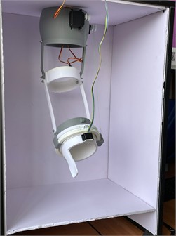
The assembled prototype is shown in Fig. 4. Assistive torque is generated by regulating cable tension, enabling passive, assistive, or active rehabilitation modes depending on the selected control strategy. The current prototype represents a preliminary structural and functional implementation intended for conceptual validation, while future work will focus on dynamic analysis, cable tension optimization, and experimental rehabilitation assessment.
Fig. 3Cable-driven mechanism scheme
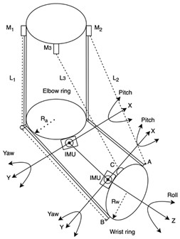
Fig. 4Experimental prototype
3. Numerical analysis and results
3.1. Model parameters and boundary conditions
Finite element analysis (FEA) was performed in SolidWorks Simulation using SI units. The exoskeleton was modeled using AISI 304 stainless steel due to its widespread use in rehabilitation and medical devices. The main material properties were: density 8000 kg/m3, Young’s modulus 1.9×10¹¹ Pa, Poisson’s ratio 0.29, and yield strength 2.07×10⁸ Pa.
A fixed boundary condition was applied to the proximal module, while external loads representing forearm motion assistance were applied to the distal segment. The analysis was conducted under simplified worst-case static loading conditions to identify critical stress concentration regions. Dynamic effects, soft tissue interaction, and cyclic loading were not considered and remain subjects for future investigation.
3.2. Finite element model
The finite element model and boundary conditions are shown in Fig. 5. A refined mesh was applied in critical regions, including joint interfaces and cable-guiding elements, to improve stress distribution accuracy. The analysis was intended as a preliminary structural assessment of the proposed concept.
3.3. Stress analysis
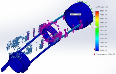
The von Mises stress distribution is presented in Fig. 6. Maximum stresses were concentrated near joint interfaces and load-transfer regions, where geometric discontinuities produced local stress concentrations 5.20×10⁹ Pa.
The obtained peak stress values exceeded the yield strength of AISI 304, indicating that the current prototype configuration requires structural refinement and reinforcement. These results highlight critical regions for future optimization rather than final mechanical performance.
Fig. 5FEM model and boundary conditions
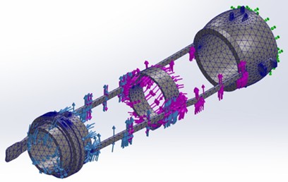
Fig. 6Stress distribution
3.4. Displacement analysis
The displacement distribution is shown in Fig. 7. The maximum displacement was observed at the distal segment: 135.5 mm.
The relatively large displacement is associated with the lightweight modular architecture and simplified boundary conditions. The results indicate limited structural stiffness under the applied loading scenario and suggest the need for further design optimization.
Fig. 7Displacement results
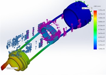
Fig. 8Strain distribution
3.5. Strain analysis
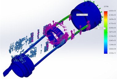
The strain distribution is presented in Fig. 8, 6.82×10⁻3.
The highest strain values occurred in regions corresponding to previously identified stress concentration zones. These areas represent critical structural locations requiring further geometric refinement and stiffness improvement.
Overall, the numerical analysis provided a preliminary assessment of the proposed concept and identified key regions requiring optimization before future experimental validation and rehabilitation testing.
4. Discussion
The numerical analysis identified several critical regions requiring structural optimization. High stress concentrations were observed near joint interfaces and load-transfer areas, while relatively large displacements occurred in the distal segment under the applied worst-case loading conditions. These results indicate that additional reinforcement and geometric refinement are necessary to improve structural stiffness and load distribution.
Despite these limitations, the proposed cable-driven architecture offers several advantages compared with conventional rigid rehabilitation systems, including remote actuator placement, reduced distal mass, improved adaptability, and reduced parasitic loading caused by mechanical-anatomical axis misalignment.
The present study represents a preliminary conceptual and structural assessment of the AlmatyExoElbow design. The analysis was limited to static loading conditions and did not include dynamic behavior, fatigue effects, cable tension modeling, or soft tissue interaction. Therefore, the obtained results should be considered as an initial evaluation intended to identify critical design regions and guide future development.
Future work will focus on structural optimization, refined numerical analysis, cable tension control, and experimental validation of the proposed rehabilitation-oriented system.
5. Conclusions
A 2-DOF cable-driven elbow exoskeleton, AlmatyExoElbow, was developed for rehabilitation applications. The proposed modular architecture improves biomechanical compatibility through remote actuator placement, reduced distal mass, and improved adaptability during elbow motion assistance.
Finite element analysis was performed as a preliminary structural assessment and identified critical stress concentration and deformation regions requiring further optimization. The obtained results indicate the need for reinforcement and refinement of several structural components.
The developed prototype demonstrates the practical implementation of the proposed cable-driven concept and provides a basis for future development. Future work will focus on structural optimization, refined numerical analysis, cable tension control, and experimental validation of the rehabilitation system.
References
-
M. G. Ceravolo and N. Christodoulou, Physical and Rehabilitation Medicine for Medical Students. Milan, Italy: Edi Ermes, 2019.
-
S. R. Anthony, P. Babu, and A. Paplikar, “Aphasia severity and factors predicting language recovery in the chronic stage of stroke,” International Journal of Language and Communication Disorders, Vol. 60, No. 3, 2025, https://doi.org/10.1111/1460-6984.70030
-
A. F. Pérez Vidal et al., “Soft exoskeletons: development, requirements, and challenges of the last decade,” Actuators, Vol. 10, No. 7, p. 166, Jul. 2021, https://doi.org/10.3390/act10070166
-
D. Duanmu, X. Li, W. Huang, and Y. Hu, “Soft finger rehabilitation exoskeleton of biomimetic dragonfly abdominal ventral muscles: center tendon pneumatic bellows actuator,” Biomimetics, Vol. 8, No. 8, p. 614, Dec. 2023, https://doi.org/10.3390/biomimetics8080614
-
M. Tian, Y. Liu, Z. Chen, X. Wang, Q. Zhang, and B. Liu, “Biomimetic design and validation of an adaptive cable-driven elbow exoskeleton inspired by the shrimp shell,” Biomimetics, Vol. 10, No. 5, p. 271, Apr. 2025, https://doi.org/10.3390/biomimetics10050271
-
Z. Chen, J. Wu, C. Ju, X. Wang, M. Tian, and B. Liu, “Design and control of a cable-driven exoskeleton system for upper-extremity rehabilitation,” IEEE Access, Vol. 12, pp. 187964–187975, Jan. 2024, https://doi.org/10.1109/access.2024.3515141
-
S. M. Sarhan, M. Z. Al-Faiz, and A. Takhakh, “EEG-based control of a 3D-printed upper limb exoskeleton for stroke rehabilitation,” International Journal of Online and Biomedical Engineering (iJOE), Vol. 20, No. 9, pp. 99–112, 2024, https://doi.org/10.3991/ijoe.v20i09.48475
-
L. J. En, N. C.-S. Ng, and R. S.-Y. Wong, “Exoskeletons in neurological rehabilitation: a commentary on current evidence and future directions,” Quantum Journal of Medical and Health Sciences, Vol. 4, No. 3, pp. 129–137, 2025, https://doi.org/10.55197/qjmhs.v4i3.147
-
A. Nasr, K. Inkol, and J. Mcphee, “Safety in wearable robotic exoskeletons: design, control, and testing guidelines,” Journal of Mechanisms and Robotics, Vol. 17, No. 5, p. 05080, 2025, https://doi.org/10.1115/1.4066900
-
T. Luecha, W. L. Yeoh, Y. Yang, J. Choi, P. Y. Loh, and S. Muraki, “Exploring grip, voice, and electromyography signals to initiate elbow flexion with a wearable robot arm,” Journal of Robotics, Vol. 2025, No. 1, p. 49882, 2025, https://doi.org/10.1155/joro/4988295
-
R. Shankar, Z. Goh, Q. Xu, E. Chew, and K. Sui Geok Chua, “Perceptions, attitudes, and lived experiences of therapists with lower limb robotic exoskeletons in stroke rehabilitation: a systematic review of qualitative studies,” Disability and Rehabilitation, pp. 1–16, 2026, https://doi.org/10.1080/09638288.2026.2637191
-
M. S. Al-Quraishi, I. Elamvazuthi, S. A. Daud, S. Parasuraman, and A. Borboni, “EEG-based control for upper and lower limb exoskeletons and prostheses: a systematic review,” Sensors, Vol. 18, No. 10, p. 3342, Oct. 2018, https://doi.org/10.3390/s18103342
-
K. I. A. Chiu et al., “Actively controlled exoskeletons show improved function and neuroplasticity compared to passive control: a systematic review,” Global Spine Journal, Vol. 15, No. 8, pp. 3933–3952, 2025, https://doi.org/10.1177/21925682251343529
-
C. B. Fidan and A. H. Akdeniz, “EEG-based brain-computer interface systems for exoskeleton control: a review with conceptual insights,” in 7th International Black Sea Modern Scientific Research, 2025.
-
A. J. Hernandez-Navarro et al., “Design, manufacturing, and electroencephalography of the Chameleon-1 helmet: technological innovation applied for diverse neurological therapies,” Applied System Innovation, Vol. 8, No. 2, p. 56, 2025, https://doi.org/10.3390/asi8020056
-
T.-Y. Kim, S.-H. Kim, and H. Ko, “Design and implementation of BCI-based intelligent upper limb rehabilitation robot system,” ACM Transactions on Internet Technology, Vol. 21, No. 3, pp. 1–17, 2021, https://doi.org/10.1145/3392115
-
S. Kotov and M. Ceccarelli, “Design and prototype of L-CADEL.v5 elbow assisting device,” Designs, Vol. 9, No. 6, p. 126, Nov. 2025, https://doi.org/10.3390/designs9060126
-
S. Martin and E. Sanchez, “Anatomy and biomechanics of the elbow joint,” Seminars in Musculoskeletal Radiology, Vol. 17, No. 5, pp. 429–436, 2013, https://doi.org/10.1055/s-0033-1361587
-
A. Alamdari and V. Krovi, “Robotic physical exercise and system (ROPES): a cable-driven robotic rehabilitation system for lower-extremity motor therapy,” in 39th Mechanisms and Robotics Conference, 2015, https://doi.org/10.1115/detc2015-46393
-
M. A. Minetto, A. Giannini, R. Mcconnell, C. Busso, G. Torre, and G. Massazza, “Common musculoskeletal disorders in the elderly: the Star Triad,” Journal of Clinical Medicine, Vol. 9, No. 4, p. 1216, 2020, https://doi.org/10.3390/jcm9041216
-
G. S. Bullock et al., “Neck range of motion prognostic factors in association with shoulder and elbow injuries in professional baseball pitchers,” Journal of Shoulder and Elbow Surgery, Vol. 34, No. 2, pp. 421–429, 2025, https://doi.org/10.1016/j.jse.2024.08.026
-
D. Bizhanov, N. Zhetenbayev, M. Ceccarelli, G. Balbayev, and K. Ozhikenov, “Design and performance of a motion assisting device for elbow joint,” in Mechanisms and Machine Science, Vol. 167, Cham: Springer Nature Switzerland, 2024, pp. 152–159, https://doi.org/10.1007/978-3-031-67569-0_18
-
D. Bizhanov, N. Zhetenbayev, G. Sergazin, A. Nussibaliyeva, S. Yussupova, and A. Maksut, “Design and simulation of a cable-driven elbow rehabilitation device,” Vibroengineering Procedia, Vol. 58, pp. 139–146, May 2025, https://doi.org/10.21595/vp.2025.24971
About this article

This research has been funded by the Ministry of Science and Higher Education of the Republic of Kazakhstan, Grant No. AP27508009.
The datasets generated during and/or analyzed during the current study are available from the corresponding author on reasonable request.
Dr. Nursultan Zhetenbayev is a scientific committee member of the 77th International Conference on Vibroengineering and was not involved in the editorial review and/or the decision to publish this article.

